This website uses cookies to ensure you get the best experience on our website.
- Table of Contents
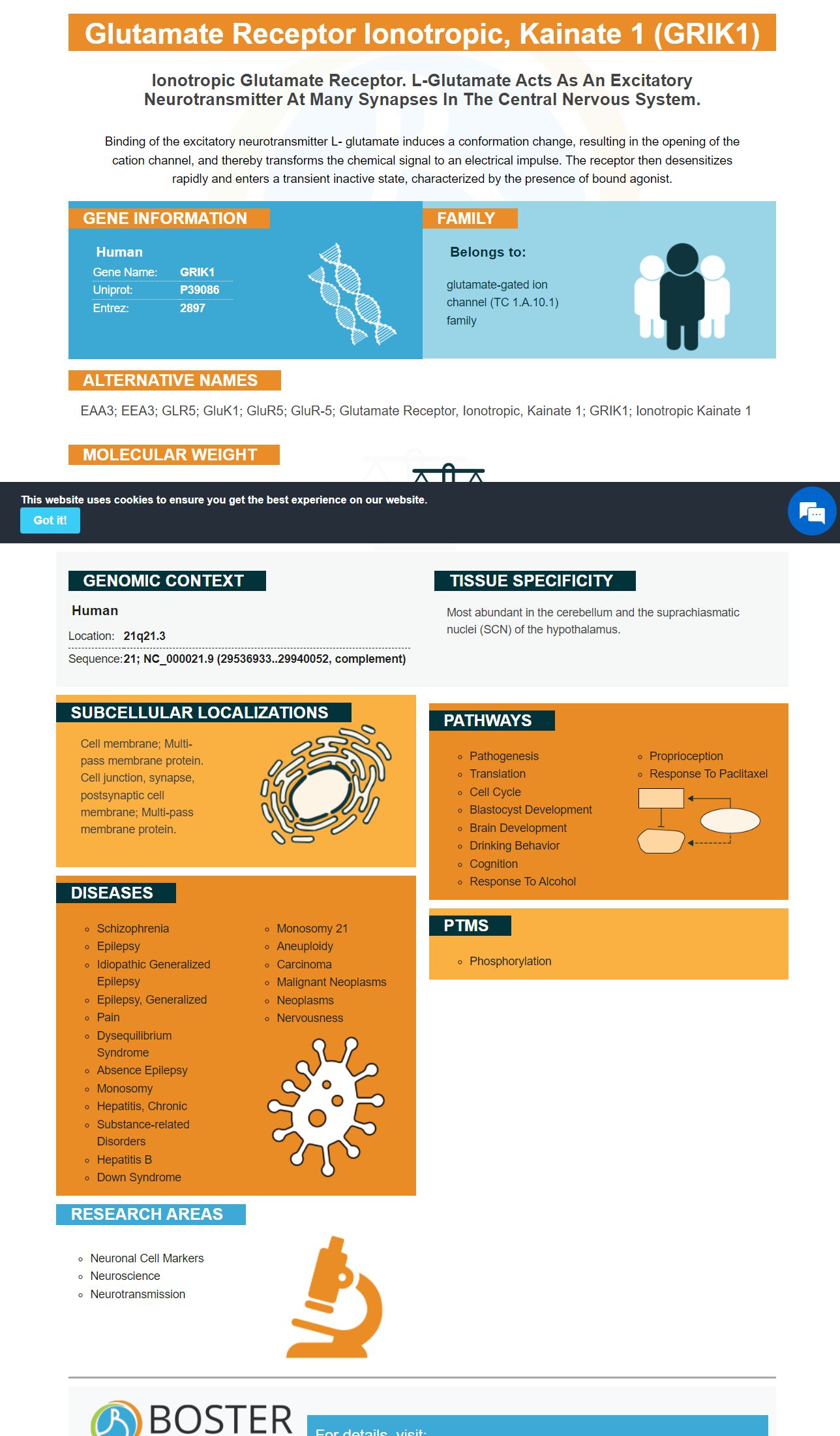
Facts about Glutamate receptor ionotropic, kainate 1.
Binding of the excitatory neurotransmitter L- glutamate induces a conformation change, resulting in the opening of the cation channel, and thereby transforms the chemical signal to an electrical impulse. The receptor then desensitizes rapidly and enters a transient inactive state, characterized by the presence of bound agonist.
Human | |
---|---|
Gene Name: | GRIK1 |
Uniprot: | P39086 |
Entrez: | 2897 |
Belongs to: |
---|
glutamate-gated ion channel (TC 1.A.10.1) family |
EAA3; EEA3; GLR5; GluK1; GluR5; GluR-5; glutamate receptor, ionotropic, kainate 1; GRIK1; ionotropic kainate 1
Mass (kDA):
103.981 kDA
Human | |
---|---|
Location: | 21q21.3 |
Sequence: | 21; NC_000021.9 (29536933..29940052, complement) |
Most abundant in the cerebellum and the suprachiasmatic nuclei (SCN) of the hypothalamus.
Cell membrane; Multi-pass membrane protein. Cell junction, synapse, postsynaptic cell membrane; Multi-pass membrane protein.
If you are interested in the Boster Bio: Anti-Grik1 Antibody, then read this article to learn more about the product and its applications. Within its second transmembrane domain, the GRIK1 Protein has many isoforms. This domain is where the protein plays a role in RNA editing. You can find out more about the functions of the protein in RNA editing.
Boster Bio Anti-Grik1 Antibodies is available in a variety reagents as well as kits. They can also be used to make whole-cell immunoblots. Boster antibodies are highly specific and affinity-specific and have been validated across multiple platforms using known positive or negative samples. Boster antibodies are also highly rewarding as they provide product credits for first-time reviewers and scientists around the globe.
To assess the efficacy of the GRIK1 marker in MS, we used RNA scope(r) Fluorescent Multiplex Kit on paraffin-embedded fresh frozen brain sections. The brains of the patients were removed with anesthesia. They were then flash frozen in dryice. The amygdala and brains were then removed and cut into 10-um-thick slices. The sections were then fixed in 4 percent PFA and dehydrated using an ethanol series. The optical density of each section was used to determine the mRNA expression. This was normalized to the number and size of the cells within the section. For each animal, a minimum of three sections were evaluated.
GRIK1+ cells had a significant decrease in density in MS rats as compared to controls. However, the number Parv+Grik1 doublepositive cell numbers showed no significant differences. Grik1 expression was decreased in non-glutamatergic neuron and parvalbumin positive interneurons in the MS amygdala. These findings suggest that GRIK1 could be useful in identifying neuronal groups that are involved a wide range of brain disorders.
GRIK1 mRNA levels in adult mice were not significantly different from those of P14 control mice. Interestingly, GRIK1 knockout mice showed a mild anxiety-like phenotype following genetic inactivation. This knockout model also affected 50% of the BLA cells, which were predominantly nonglutamatergic. Furthermore, the expression of GRIK1 was significantly downregulated in mice with male sex. The results showed that GRIK1 expression was significantly downregulated in mice following ELS.
GluK1 regulates PV+ neuron excitability, which in turn gate AP firing of SOM+ neurons through inhibitory GABAergic connection. KAR-dependent disinhibition in principal cells during afferent activation may be explained by the shift in excitability between SOM+ and PV+ interneurons. These studies are not conclusive and more research is needed to determine if GRIK1 plays a role in neuronal excitability.
Multiple GRIK1 isoforms are possible to be identified. Its second transmembrane domain is made up of two subunits: GRIK1 (or GRIK2) and GRIK2. These subunits have different abilities to regulate ion flow. GRIK1 expression is also found in tumors and cells. This suggests that GRIK1 might be involved with the regulation of EMT.
The GRIK1 gene is composed of four isoforms. GluK1a and GluK1b share a similar splicing patterns. GRIK1b's GluK1b is expressed at a tenfold less level. Its global abundance is modestly higher than that of GluK4b, which has a 6% abundance in humans. GRIK1b-b has an unusual event called exon skipping in human samples.
There is no consensus on the exact biological role of GRIK1. Its function remains unclear. While there is a high probability that GRIK1 plays a role in tumor suppression in vivo, it is unclear whether GRIK1 has any such function. However, Minbay et al. reported that heteromeric functional receptor channels were formed by combining different ionotropic glutamate receptor subunits in the red nucleus neurons of adult female Sprague-Dawley rats. To determine if GRIK1 has tumor suppressor properties, further functional and pharmacological research is needed.
The two FACS methods used for identifying the BC2 - BC4 subtype markers are highly compatible. Grik1CRM4 reporter vector plasmids allowed us to determine relative expression levels of each subtype markers gene by scRNA sequencing. ScRNA sequencing enabled us to identify BC2-BC4 marker genes as well as specific cell classes and isoforms.
IHC staining on 80 CRC cells was done. The expression levels of GRIK1 were graded as weak, moderate, or strong, based on their expression. Tumor size and lymphovascular invasion were associated with low GRIK1 expression. This study supports the idea of GRIK1 being a useful marker for CRC detection. This could help to identify these tumor cells using immunohistochemical methods.
Although RNA editing in the second transmembranes domain has been described before, it was not widely reported. This study reveals how the ADAR2 protein regulates RNA edits at this site. ADAR2 modifies the GluA2Q/R site in a primary way, and ADAR3 expression is restricted to the brain. Although ADAR3's exact role remains unknown, it may regulate RNA edit at this site.
GRIK1 marks allow for RNA editing to alter the ion flows properties of the cell membrane. Within the second transmembrane domain, we found multiple GRIK1 isoforms. Some of these areoforms were enriched simultaneously while others had low or moderate abundance. These rare events are listed on Table S4.
Highly edited reads include GluK2Q621R/G615G. This marker is highly expressed non-neuronal tissue, and a substantial percentage of them were identified by de Novo analysis. However, reads edited by the GluK2Q621R/G615G website were not overrepresented in this data.
Evidence indicates that RNA editing at the Q/R site is essential for survival and neuronal function. This editing does not stop the growth of Ca2+ permeable AMPA receptors. Unedited GluA2 protein could play a role as a neuronal loss marker in certain diseases. However, it is not known if this is essential.
Cell survival is improved by blocking Ca2+-permeable AMPA receivers. Further studies are needed to determine if unedited GluA2 contributes in any way to cell survival in diseases. Electrophysiology, in addition to the RNA editing method, may be used to determine if functional unedit GluA2 mRNA exists.
GluK1-a isoforms are more likely to be expressed in heterologous expression systems. Although most functional data was obtained with GluK1a, the RNA editing genes also has known splicing variations. The first variant involves preserving signal peptide by skipping exon 2. 56 amino acids are removed from the ATD's N-terminal section in the second variant.
PMID: 8260617 by Gregor P., et al. Expression and novel subunit isoforms of glutamate receptor genes GluR5 and GluR6.
PMID: 8589992 by Korczak B., et al. cDNA cloning and functional properties of human glutamate receptor EAA3 (GluR5) in homomeric and heteromeric configuration.