This website uses cookies to ensure you get the best experience on our website.
- Table of Contents
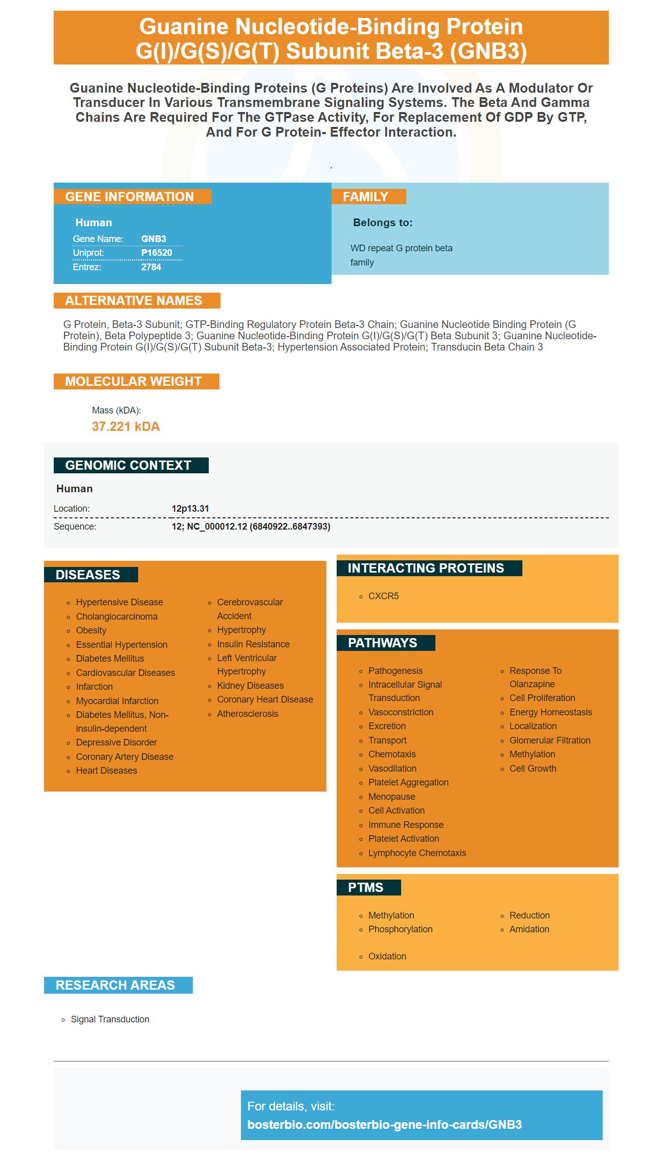
Facts about Guanine nucleotide-binding protein G(I)/G(S)/G(T) subunit beta-3.

.
| Human | |
|---|---|
| Gene Name: | GNB3 |
| Uniprot: | P16520 |
| Entrez: | 2784 |

| Belongs to: |
|---|
| WD repeat G protein beta family |

G protein, beta-3 subunit; GTP-binding regulatory protein beta-3 chain; guanine nucleotide binding protein (G protein), beta polypeptide 3; guanine nucleotide-binding protein G(I)/G(S)/G(T) beta subunit 3; guanine nucleotide-binding protein G(I)/G(S)/G(T) subunit beta-3; hypertension associated protein; Transducin beta chain 3
Mass (kDA):
37.221 kDA

| Human | |
|---|---|
| Location: | 12p13.31 |
| Sequence: | 12; NC_000012.12 (6840922..6847393) |


The GNB3 marker is a human protein produced by E.coli. It contains a His-Tag and a sequence of one hundred and thirty-four amino acids. It is useful for SDS-PAGE and other denatured applications. It can be stored at 2degC to eight degrees Celsius for a week. Alternatively, it can be stored at -20degC to eighty degrees Celsius for up to three months. However, it should not be subjected to repeated freeze-thai cycles. You can also earn credits for reviewing the product. You can even earn product credits if you are the first to review it. This opportunity is open to scientists around the world.
The choice of antibody detection method is usually determined by the level of antigen expression. For instance, the use of a primary antibody directly conjugated to a label provides a simple method of multicolor staining. However, direct detection lacks sensitivity when used to visualize antigens of low expression levels, and the conjugation process can negatively impact the affinity of the primary antibody. Listed below are the main types of antibody detection methods that use the GNB3 marker.
The GNB3 protein is expressed in a subset of bipolar cells in the human body, which is also highly conserved in many species. The GNB3 C825T polymorphism results in aberrant splicing in approximately 50% of the time. As a result, both normal Gb3 proteins and truncated Gb3s are produced. Previous studies have linked the 825T allele with obesity, hypertension, various cancers, and age-related cognitive function.
The GNB3 C825T SNP has been associated with HIV-1 acquisition, disease progression, and the prevalence of HIV-1 in Africa. Further research is needed to examine its effects in non-African populations and its mechanism of action. Until then, antibody detection methods using the GNB3 marker remain an effective method for detecting HIV-1 in blood samples. So, why not give it a try?
To create high-affinity primary antibodies using the GNB3/GNB3 marker, the first step is to purify and lyse 105 hPDLSCs in a 6-well tissue culture plate. Next, specific antibodies were conjugated with allophycocyanin, fluorescein isothiocyanate, or phycoerythrin-cyanine 5.5. After conjugation, hPDLSCs were stained with Ancell.
Conjugated primary antibodies are ideal for applications requiring high specificity and affinity. These antibodies are highly useful in immunoblotting, ELISA, immunofluorescence microscopy, flow cytometry, and immunohistochemistry. High-affinity primary antibodies can be used for most immunological techniques. The GNB3 marker is particularly useful for this application. These antibodies are suitable for all immunological applications, including the detection of proteins that may cause disease.
To measure KD, the antibody solution was injected at time zero and ran across the peptide microarray for 15 min. The dissociation reaction was then calculated by comparing the on-rate and off-rate of each antibody/antigen complex. The lower the KD, the greater the affinity of the antibody. This data was evaluated by scientists from Abcam and UC Davis.
The KD distribution of RabMAbs differs from that of mouse MAbs. Mouse MAb KD values were derived from published literature. In contrast, RabMAb antibodies were obtained from Ol-RD measurements. The median KD for RabMAb antibodies was 7 x 10-11 M, which is an excellent correlation with the other methods of analysis. The results of this study show that GNB3-based primary antibodies have high affinity and sensitivity.
A Western blot is a specialized method for detecting protein expression in cells and tissues. It uses antibodies to house-keeping proteins which thiexpressed at comparable levels in all cells and tissues. The quality of the blot depends on the method of sample preparation and protein extraction. Diagenode's H3pan monoclonal antibody is a useful loading control for nuclear samples. It helps compare protein expression levels between two samples.
The Diagenode antibody was used to perform the western blot. This antibody was diluted 1:4,000 in PBS-T containing 3% NFDM. The Western blot shows the position of the Cas9 protein and marker. The marker was present on the left blot as indicated on the right. Generally, a protein is detected if its band is visible.
This technique can detect protein levels as low as 1ng. The high resolution of gel electrophoresis allows for detection of such low levels. This sensitivity and specificity of the immunoassay make it an essential part of molecular biology. The Western blot method is used in biochemistry, immunogenetics, and molecular biology. The GNB3 marker can be detected in a variety of biological samples.
In addition, the antibody is also available as fluorophore-conjugated antibodies. The latter is the preferred method, as it requires fewer steps and does not require expensive special equipment. The fluorophore-conjugated antibodies thialso more expensive, but the advantages of using them over monoclonal antibodies include their sensitivity, multiplex compatibility, and reduced chemical waste.
The ECL chemiluminescent detection (ECL) technique is more sensitive than traditional colorimetric assays. ECL is a highly sensitive detection method that detects proteins with low-to-medium levels of expression. The ECL system uses Luminol, a molecule with a short half-life of a few minutes. Its sensitivity is up to 1000-fold higher than Luminol, and its enhanced chemiluminescence (ECL) reagents can detect medium to-high-expression levels of protein.
The method begins with a primary antibody that recognizes the target protein and a secondary antibody that is conjugated to an enzyme. A typical enzyme used in ECL detection is horseradish peroxidase (HRP), alkaline phosphatase (AP), or luminol-based HRP. Once the reaction begins, light photons are measured using an x-ray film or digital imaging.
A high-salt wash buffer, pH 7.4, contains 10 mM Tris, 0.5 M NaCl, 1 mM NaH2PO4, 0.2% SDS, and 1% Tween-20. The Boster BLoT Ultra Sensitive HRP Substrate is highly sensitive, allowing for the detection of very low levels of protein or antigen. High-salt aqueous buffer is compatible with most nitrocellulose membranes and produces very low background levels. The high salt aqueous solution provides a clear and linear emission signal that is highly visible on CCD imaging systems.
A C-DiGit scanner eliminates the need for a darkroom, developing reagents, and film. The Odyssey XF is another option that offers chemiluminescent protocols and full NIR imaging. This system can be used for Western blots, plate-based assays, and excised organs. This method is suitable for high-throughput experiments where the presence of protein is important.
DAB chromogenic detection is a critical step in the diagnosis of various diseases. The system uses a dye known as 3,3'-diaminobenzidine (DAB) to detect the presence of the marker. In previous studies, the main focus was on determining the activity of AR, but localization and visual expression were not reported. Most of the studies detected TLR4 by using conventional immunohistochemistry, which is well-established for tissue and cell imaging. The DAB staining intensity depends on the horseradish peroxidase substrate, and the reaction time will affect the quantitative detection sensitivity.
A Western blotting protocol is an important step in the process of analyzing protein expression. Typically, this process consists of two steps: SDS-polyacrylamide gel electrophoresis and protein blotting. The steps for the Western blotting protocol can vary slightly depending on the reagent used, but the basic procedure involves incubation with a protein-specific secondary antibody and membrane staining. The use of a Western blotting protocol can help reduce the possibility of artifacts. Artifacts in the western blotting protocol can be caused by uneven coating or aggregates of the antibody.
One of the most popular methods for staining proteins is Coomassie Brilliant Blue. It is inexpensive and easy to visualize. The Coomassie Brilliant Blue binds proteins non-covalently via hydrophobic and ionic interactions. After staining, bands of Coomassie Brilliant Blue-stained proteins appear as dark blue bands on light blue backgrounds. This method also allows researchers to monitor protein transfer efficiency and identify alterations in protein concentration.
For this method, diluted healthy mixed sera was separated by 8% SDS-PAGE, and then transferred to a PVDF membrane. Afterward, the proteins were re-probed using an antibody, lectin, or both. The antibodies were able to detect the target proteins, and this method allows for more accurate protein transfer. Once the antibodies bind the target protein, the membrane can be used for subsequent antibody staining.
PMID: 2107550 by Levine M.A., et al. Molecular cloning of beta 3 subunit, a third form of the G protein beta-subunit polypeptide.
PMID: 8723724 by Ansari-Lari M.A., et al. A gene-rich cluster between the CD4 and triosephosphate isomerase genes at human chromosome 12p13.


