This website uses cookies to ensure you get the best experience on our website.
- Table of Contents
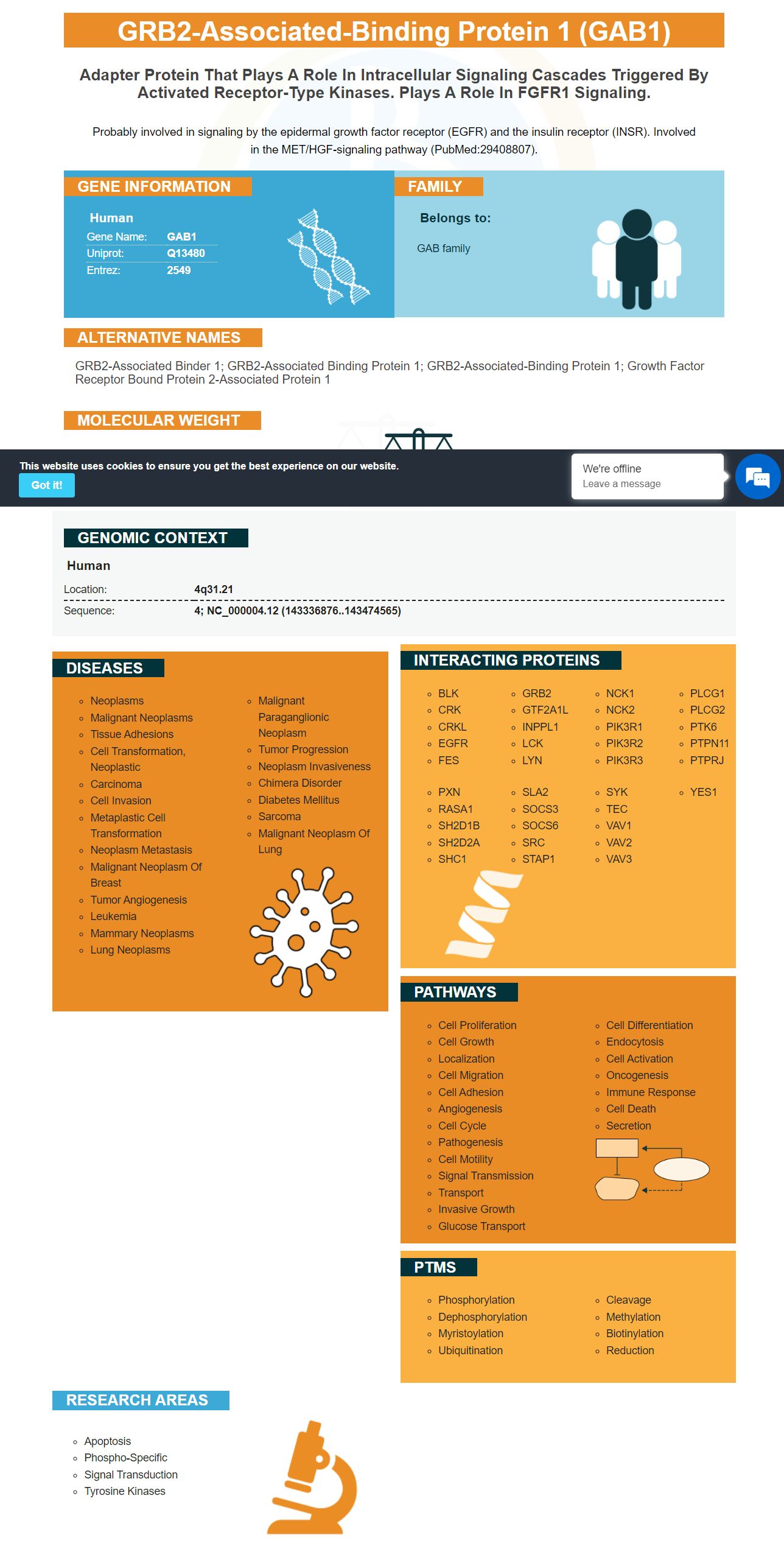
Facts about GRB2-associated-binding protein 1.
Probably involved in signaling by the epidermal growth factor receptor (EGFR) and the insulin receptor (INSR). Involved in the MET/HGF-signaling pathway (PubMed:29408807).
Human | |
---|---|
Gene Name: | GAB1 |
Uniprot: | Q13480 |
Entrez: | 2549 |
Belongs to: |
---|
GAB family |
GRB2-associated binder 1; GRB2-associated binding protein 1; GRB2-associated-binding protein 1; Growth factor receptor bound protein 2-associated protein 1
Mass (kDA):
76.616 kDA
Human | |
---|---|
Location: | 4q31.21 |
Sequence: | 4; NC_000004.12 (143336876..143474565) |
A Boster scientist can submit his or her results for species, applications, and special samples. By submitting his or her results, scientists can get product credits and be recognized in the Boster Bio database. This program is open to scientists from any country worldwide. Read on to learn more about Boster Bio: Best Uses Of The GAB1 Marker
Ki-67, or KIA, is a polypeptide encoded by the MKI67 gene. Schonk et al. identified this gene on chromosome 10 by using a panel of rodent-human somatic cell hybrids. Later, Fonatsch et al. localized the human MKI67 gene to chromosome 10q25-qter. Similarly, Traut et al. mapped the mouse MKI67 gene to chromosome 7F3-F5 in 1998. However, despite the high concentration of anti-Ki67 antibodies, it is unclear whether this antibody is effective in detecting this marker in tumor cells.
In the study presented here, human Jurkat cells were fixed, permeabilized, and stained with Anti-AKT pS473 and REA Control (I) antibodies. The cells were analyzed by flow cytometry using a MACSQuant(r) Analyzer. To increase the binding specificity of tandem-dye-conjugated antibodies, Tandem Signal Enhancer was used. Moreover, cell debris was excluded from the analysis based on scatter signals.
Monoclonal antibody to phosphorylated human and mouse AKT was produced by repeated immunization of mice with a synthetic peptide. Antibodies are purified by Protein A chromatography. This monoclonal antibody recognizes phosphorylated human and mouse AKT1.
The primary antibody clone 14-5 recognizes Akt phosphorylated at S473. The phosphorylation site is found on all three isoforms of Akt. Antibodies to Akt2 and Akt3 recognize homologous phosphorylation sites. Activated Akt is activated in various types of cancer, including prostate, lung, breast, and renal cancer.
The antibody was tested against a known anti-Akt antibody from Cell Signaling. The Rockland antibody failed to detect phosphorylated Akt. The Cell Signaling antibody showed that activated Akt was phosphorylated when exposed to HGF. These findings confirm that the rabbit anti-Akt antibody can detect phosphorylated Akt in cancer cells. A rabbit anti-AKT pS473 (MOUSE) antibody can detect phosphorylated Akt in human breast cancer tissue.
In immunohistochemistry, Phospho-AKT antibody recognizes endogenous levels of human AKT1 phosphorylated at Ser473. It does not recognize phosphorylated forms of related kinases. It is produced by immunizing mice with synthetic phosphopeptide. Afterwards, peptide affinity chromatography is used to purify polyclonal antibodies.
Akt, also known as RAS-alpha, phosphorylates several proteins in the cell. It is activated by various growth factors, insulin, and PI3K. It inhibits apoptosis and promotes proliferation. As such, Anti-AKT antibodies are an excellent choice for Neuroscience and Cell Signaling research. So, what's next? Take your pick!
PMID: 8596638 by Holgado-Madruga M., et al. A Grb2-associated docking protein in EGF- and insulin-receptor signalling.
PMID: 12486104 by Brdicka T., et al. Non-T cell activation linker (NTAL): a transmembrane adaptor protein involved in immunoreceptor signaling.