This website uses cookies to ensure you get the best experience on our website.
- Table of Contents
Facts about Follitropin subunit beta.
Human | |
---|---|
Gene Name: | FSHB |
Uniprot: | P01225 |
Entrez: | 2488 |
Belongs to: |
---|
glycoprotein hormones subunit beta family |
follicle stimulating hormone, beta polypeptide; Follicle-stimulating hormone beta subunit; Follitropin beta chain; follitropin subunit beta; Follitropin; follitropin, beta chain; FSH beta; FSHB; FSH-B; FSH-beta
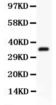
Mass (kDA):
14.7 kDA
Human | |
---|---|
Location: | 11p14.1 |
Sequence: | 11; NC_000011.10 (30231014..30235277) |
Secreted. Efficient secretion requires dimerization with CGA.
FSHB is a transmembrane receptor that binds to a number of biomolecules. Scientists are using this biomarker to analyze a variety of biological samples, including species and applications. Scientists can submit their results to receive product credits or submit special samples for their work. The program is open to all scientists from around the world, and the use of this biomarker is not limited to the U.S. military.
The antiserum against the FSHB is available as a catalog number PB10065. It has been tested for WB applications and can be stored at -20°C or 4°C for up to one year. The product contains 5mg of BSA. The antibody is available for purchase at a cost of $49 per gram. It is also applicable to other scientists from around the world.
FSHR is a transmembrane receptor with a seven-helical structure. FSH binds to its intracellular domain and activates it, by binding a G protein bound to it. Once it binds with FSH, the G protein detaches from the receptor and activates the cAMP system. The G protein's signal chain activates protein kinases.
The FSHR is a transmembrane receptor that interacts with follicle-stimulating hormone. It represents the family of G protein-coupled receptors. The receptor binds with the hormone in the ovary and testis. It contains seven membrane-spanning domains and serine and threonine residues. Hence, the FSHR has an important role in mammalian reproduction.
The FSHB is an important mediator for retinol transport and is essential for the differentiation of ovarian granulosa cells. This hormone plays a role in female fertility. If it can be induced in ovarian follicles, the hormone will influence the production of ovaries. However, in ovarian cancer patients, the FSHB is not expressed in ovaries.
FSHR is an autosomal recessive polycystic ovary disorder caused by mutations in the FSHR gene. This gene encodes a protein that binds to a transmembrane receptor on ovarian granulosa cells, resulting in the production of estradiol. Approximately half of affected females have primary amenorrhea and only one-half have breast development. Some females may have normal breast development and initiate menstrual periods. In contrast, a mutation in the FSHR gene is not as severe, but does affect binding and signal transduction.
FSHR is a protein with nine small exons. Nine of these encode the extracellular ligand-binding domain, while the remaining three code for a seven-transmembrane domain and an intracellular domain. The Finnish mutation, located in the extracellular domain, is a substitution of Ala1989Val. Mutant cells expressed with this mutation displayed a very small FSH-induced effect on cAMP production, but normal binding affinity.
Moreover, the FSHB binding ectodomain is essential for efficient receptor activation by hormones. The hinge domain is found to harbor an epitope and is responsible for transmembrane ligand binding. Moreover, monoclonal antibodies targeting FSHB or FSH can suppress basal activity. But the results of this work cannot be interpreted as proof that the FSHB binds to a transmembrane receptor.
Studies have revealed that FSHR oligomerization is a constitutive process. These oligomers are formed by hydrophobic interactions between LRR 2-4 and the exodomain of FSHR. These oligomers are found in the ER where they are involved in biosynthesis. The discovery of FSHR oligomers by Mazurkiewicz et al. helped shed light on the regulation of signaling in FSHR.
In addition to recognizing a specific antigen, FSHB Marker is also a highly efficient immunogen that can improve antibody affinity by up to 10 thousand-fold during immune response. High affinity antibodies strengthen the immune response by binding to the antigen and inhibiting its growth. Besides, the high-affinity marker is a very useful diagnostic tool for various diseases. It can be applied to detect autoimmune diseases and other inflammatory conditions.
The FSHB Marker is a high affinity primary antibody that is produced through a process that involves multiple steps and a reagent. The primary antibody recognizes the target protein and the secondary antibody amplifies the signal and enables multiple detection methods. The selection of the primary antibody and the secondary antibody depends on a number of factors. For instance, if the target protein is Strep-tagged, a murine StrepMAB-Classic antibody would be suitable.
The affinity of a single molecule is usually measured as the equilibrium dissociation constant, or KD. This KD value is an inverse relationship between the concentration of the target protein and the affinity of the antibody. As affinity is a reversible process, the rate of affinity binding equals the rate of dissociation into its constituent components. Various reactions have been carried out to determine the KD (Kelch-Dissociation constant). When comparing antibodies of the same affinity, the lower KD value corresponds to higher affinity.
Both antibodies contain insertions in VHFR3 which allow the antibodies to extend between glycans and under the glycan shield. In addition to fshb, the antibodies also recognize the FSHBP marker. The insertions in VHFR3 allow them to detect gp120 and its epitope. If these three regions overlap, the corresponding epitopes will be marked by red and blue sticks.
The FSHB marker is a protein found in human cells. Induction of FSHB is achieved through activin. Transfection of IGSF1 represses FSHB transcriptional activity. Activin stimulates expression of FSHB, but IGSF1 does not. Therefore, IGSF1 is a strong repressor of the activin pathway. Its basal transcriptional activity is reduced by 50% when co-transfected with CAGA.
To use the FSHB marker in IHC, formalin-fixed tissues must be boiled in 10mM Citrate buffer, pH 6.0. Tissue sections should be cooled to room temperature (RT) after the boiling. The prediluted FSHB marker is available in a dropper bottle. Its dilution is optimized for IHC and epitope retrieval, and the incubation time is 30 minutes.
FSHR323 antibody recognizes a specific epitope on tumor endothelial cells. FSHR190 and FSHR225 monoclonal antibodies recognize the same epitope and have similar staining patterns. The FSHR323 antibody is not specific for prostate cancer, and the FSHR18 monoclonal antibody recognizes a different epitope.
Several studies have demonstrated that the FSHB marker is specific for the adenopituitary lobe and the adenopituitary. Using a nonimmune mouse IgG2a antibody can distinguish between tumors and normal tissue. Another study used the FSHB marker to differentiate between benign and malignant tumors. In addition, FSHB can help define the timing of stimulation of the Sertoli cell in IGSF1 deficiency.
FSHB is a common human hepatitis B virus antigen that is found in the urine of patients. The ELISA technique is used to detect antibodies that detect this hepatitis B virus antigen. It is a sandwich assay in which cells are plated and proteins are detected by a precipitating substrate. Like the western blot, ELISPOT produces spots on the membrane surface.
An ELISA involves the use of a reagent that binds to the target protein. It uses a marker called FSHB to determine the presence of a specific antigen. ELISA is a sensitive, rapid method for detecting antigens. The ELISA is highly sensitive, and can detect a single molecule in just three hours. The ELISA method is a sensitive and accurate test that can identify several antigens.
ELISA is an excellent tool for measuring specific analytes in crude preparations. The high-affinity antibodies used in ELISA prevent nonspecific binding and make it an efficient method for measuring specific analytes in a crude preparation. Hence, the ELISA is a cost-effective method for detecting antigens in a variety of samples. The FSHB marker is one of the most popular and widely used protein markers.
Another benefit of FSHB marker in ELISA is the possibility to measure gonadotropin bioactivity in a single sample. Currently, a microELISA has been developed that can measure nine different antigens in a single sample. This multiplexed sandwich ELISA has been validated in 44 clinical samples. In addition to the FSHB antigen, it can measure other proteins, such as adenosine and insulin.
PMID: 2885163 by Watkins P.C., et al. DNA sequence and regional assignment of the human follicle- stimulating hormone beta-subunit gene to the short arm of human chromosome 11.
PMID: 2494176 by Keene J.L., et al. Expression of biologically active human follitropin in Chinese hamster ovary cells.