This website uses cookies to ensure you get the best experience on our website.
- Table of Contents
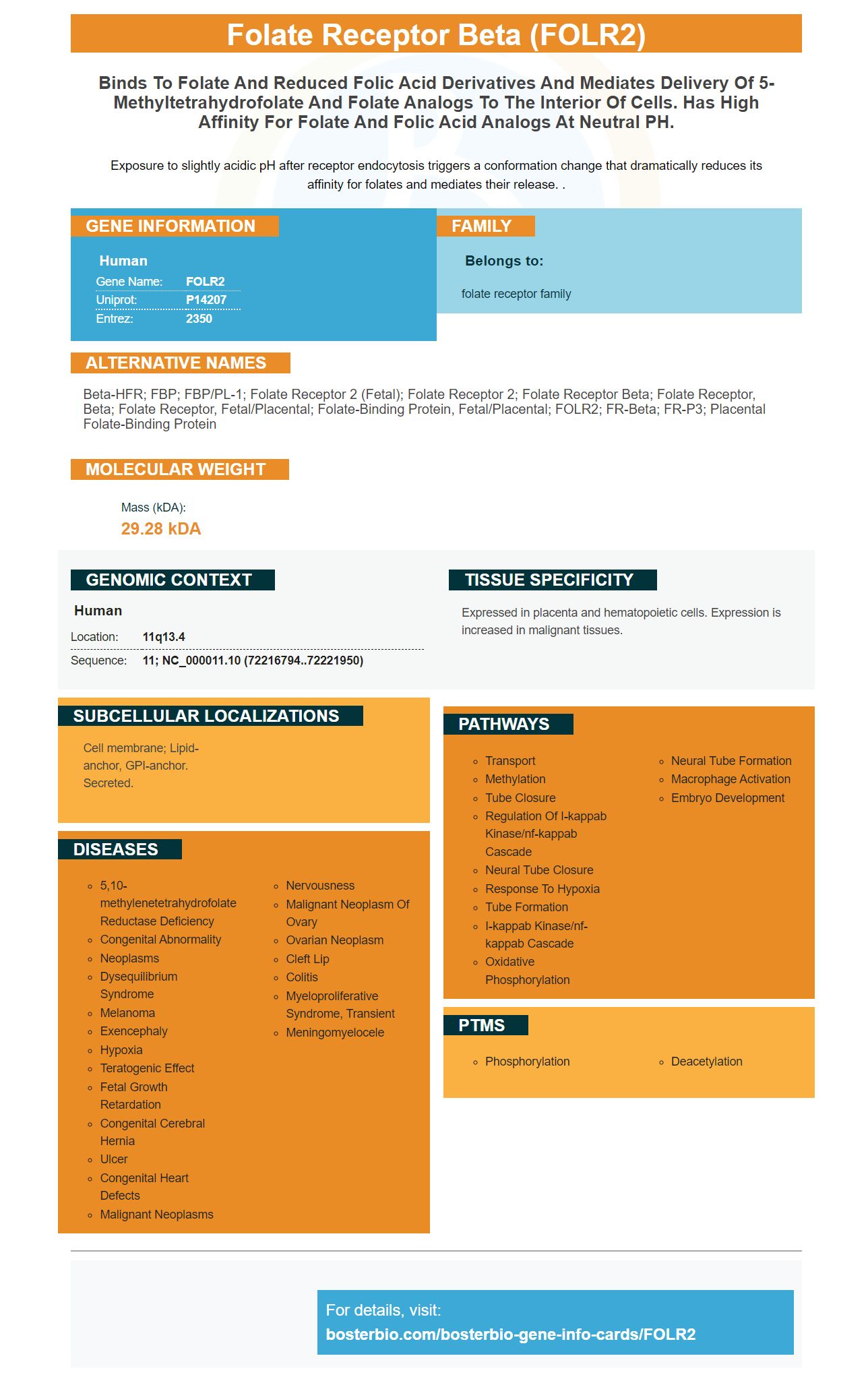
Facts about Folate receptor beta.
Exposure to slightly acidic pH after receptor endocytosis triggers a conformation change that dramatically reduces its affinity for folates and mediates their release. .
Human | |
---|---|
Gene Name: | FOLR2 |
Uniprot: | P14207 |
Entrez: | 2350 |
Belongs to: |
---|
folate receptor family |
beta-HFR; FBP; FBP/PL-1; folate receptor 2 (fetal); Folate receptor 2; folate receptor beta; folate receptor, beta; Folate receptor, fetal/placental; folate-binding protein, fetal/placental; FOLR2; FR-beta; FR-P3; Placental folate-binding protein
Mass (kDA):
29.28 kDA
Human | |
---|---|
Location: | 11q13.4 |
Sequence: | 11; NC_000011.10 (72216794..72221950) |
Expressed in placenta and hematopoietic cells. Expression is increased in malignant tissues.
Cell membrane; Lipid-anchor, GPI-anchor. Secreted.
Boster Bio's products are a reliable source of FOLR2-recognising antibodies. Its primary antibodies are routinely validated on WB, IHC, and ICC, and have been proven effective in a range of applications. Boster also rewards the first reviewers with product credits, making their antibodies a great choice for researchers from different disciplines.
Boster has been producing reliable, high affinity antibodies for ELISA and Western Blotting for over 25-years. Their primary antibodies are highly regarded in the immunology community and have been extensively validated in Western Blotting, Immunohistochemistry, and ELISA. Boster's quality products and commitment to quality have earned them the trust of researchers across the globe.
Their antibodies are unmatched in quality. Boster is an ISO 9001 accredited company. Researchers across the world rely on their primary antibodies to perform experiments in immunocytochemistry, cell biology, and genetics. Their antibodies are a valuable component in research. Because they are 100% human, researchers trust them. The company has an excellent reputation for quality and reliability, and its products are highly priced, affordable, and widely available.
Rockland's primary antibodies are versatile. They recognize antigens that have high affinity and specificity. The antibodies are unconjugated by default, but are often conjugated to eliminate the need for secondary antibodies. You can use conjugated primary antibodies without the need for secondary antibodies or additional chemicals, depending on your assay requirements. These antibodies can also be used in Western Blotting and other immunological applications.
Developed by scientists with years of experience, SouthernBiotech produces high-quality monoclonal antibodies, rnai, and crispr ko products for research and diagnostics. With the help of their highly-trained staff, SouthernBiotech products help researchers understand how the body processes the antigen. It is also committed towards meeting the highest industry standards. Boster's company is smaller, but their quality is exceptional.
Boster Bio, a renowned antibody manufacturer, focuses on producing IHC-optimized polyclonal antibodies and picogram-sensitive ELISA kits. Their extensive array of antibodies includes highly-specific ELISA kits as well as WB/IHC compatibility antibodies. The company has been involved in the development of their techniques for over two decades and has been referenced by more than 29,000 scientific papers. Boster Bio has high-quality antibodies and is certified by WB, IHC & Flow Cytometry.
To determine whether an antibody is specific, two antibodies to the exact same protein are tested against one another. A reagent that binds to two distinct regions of the same protein is used to compare the staining patterns of two different antibodies. Antibodies which produce similar staining patterns have to be specific and nonoverlapping. You can also verify binding by comparing the signals produced by over-expressed and tagged target proteins. Boster's antibodies are highly reliable in both IHC as well as WB applications.
The Boster Bio lab produces quality reagents and kits. Their extensive product portfolio includes buffers, assay kits, and lysates. Boster produces antibodies as well as a wide variety of controls, solutions, and lysates. These reagents can be used in many research and diagnostic applications. The company uses ELISA testing and limiting dilution cloning to verify the antibodies are as specific as possible during antibody development.
WB is, as I mentioned, an important validation tool. Knockouts also offer valuable validation tools. These methods come with limitations that must be considered. Both methods are useful in determining if a monoclonal is really specific. Negative controls for IHC or WB must be considered as they can detect false negatives and other nonspecific binding.
Boster's antibodies are also validated on tissue microarrays (TMA) along with ICC and WB. TMA is a widely-used immunogen because it allows you to identify and quantify the target. While TMA may not be a substitute for IHC, a good antibody will only stain target cells and will decrease with increasing dilutions.
Recently, the FOLR2 marker, which is a protein expressed by macrophages, was identified. The duplex RNAscope was used to examine the expression of FOLR2 within these cells. It also identified other markers, CD68b and CD11b. Some macrophages express both markers, while others only one. CD68 and FOLR2 are expressed in both cell type, but the former is more compatible with the expression than the latter.
RNAscope, a unique tool for chromogenic ICC hybridization (ICC), was used as a tool to determine the folate receptor expression in a variety os tissues and cell types. FOLR1 expression was found in normal fallopian tube, ovarian epithelial and other cells. FOLR2 expression is rare. In addition, the co-expression of FOLR2 and CD11b was found to indicate the presence of macrophages in the samples.
In addition to the FOLR2 marker, there is also a FRA or FRB. These cells are inflammatory cells and can promote tumor development and metastasis. FOLR2 expression in gynecologic tissues was evaluated using macrophage markers, such as CD11b and CD68, as well as FOLR2 probes. FOLR1 & FOLR2 were found in scattered cells, aggregating cells, and low-expressing ones in the fallopian tubes.
FOLR2(folate receptor beta), is one of the markers linked to tumors. This protein is found in macrophages that have M1 and M2 phenotypes. FOLR2 is also an indicator for cancer infiltrating microphages. These cells contain the folate-receptors, which are associated with disease progression.
The most powerful immunochemical technique for detecting many antigens is immunofluorescence. Immunofluorescence offers many advantages over traditional histology. It has high sensitivity, amplifies the signal and can be used in a variety microscopy techniques. There are two basic methods of immunofluorescence staining currently available. These differ in terms of the experiment's scope and the types of antibodies used. We'll be discussing both methods in detail.
Direct immunofluorescence uses an anti-IgA antibody to visualize the IgA deposits in the walls of small superficial capillaries. The epidermis is located on the top of the skin. While the fibrous tissue is at the bottom, it is the dermis. This technique is versatile and sensitive, making it an invaluable diagnostic tool for many diseases. Anti-IgA antibodies are useful in diagnosing autoimmune diseases and other inflammatory conditions.
The primary function of immunofluorescence (or immunofluorescence) is to visualize specific proteins or molecules in a sample. This is done using antigens and antibodies in an specialized method called immunofluorescence. This technique involves tagging specific antibodies with fluorescent dyes, which allows for visualizing the target molecule in a cell under a light microscope. A good antibody is essential for immunofluorescence to work. The antibodies must recognize antigens in cells for the technique's to work correctly.
Secondary antibodies are antibodies that are conjugated to a variety of labels, including fluorescent dyes. These antibodies can enhance the fluorescent signal if they bind directly to a primary antigen. Secondary antibodies can also help in immunofluorescence, such as diagnosing certain diseases. They can also bind to a variety different fluorophores. This method allows for a rapid, easy and accurate test of a large number of bacteria and other pathogens.
Primary immunofluorescence, which has a single antibody conjugated to a fluorophore, is a much simpler procedure that utilizes just one antibody. The primary antibody recognizes the target molecule, while the secondary antibody carries the fluorescent dye and is detected with the help of a microscope. Primary immunofluorescence has a lower number and greater sensitivity than indirect immunofluorescence.
PMID: 8445646 by Page S.T., et al. Expression of the human placental folate receptor transcript is regulated in human tissues. Organization and full nucleotide sequence of the gene.
PMID: 2605182 by Ratnam M., et al. Homologous membrane folate binding proteins in human placenta: cloning and sequence of a cDNA.