This website uses cookies to ensure you get the best experience on our website.
- Table of Contents
1 Citations 7 Q&As
Facts about Folate receptor alpha.
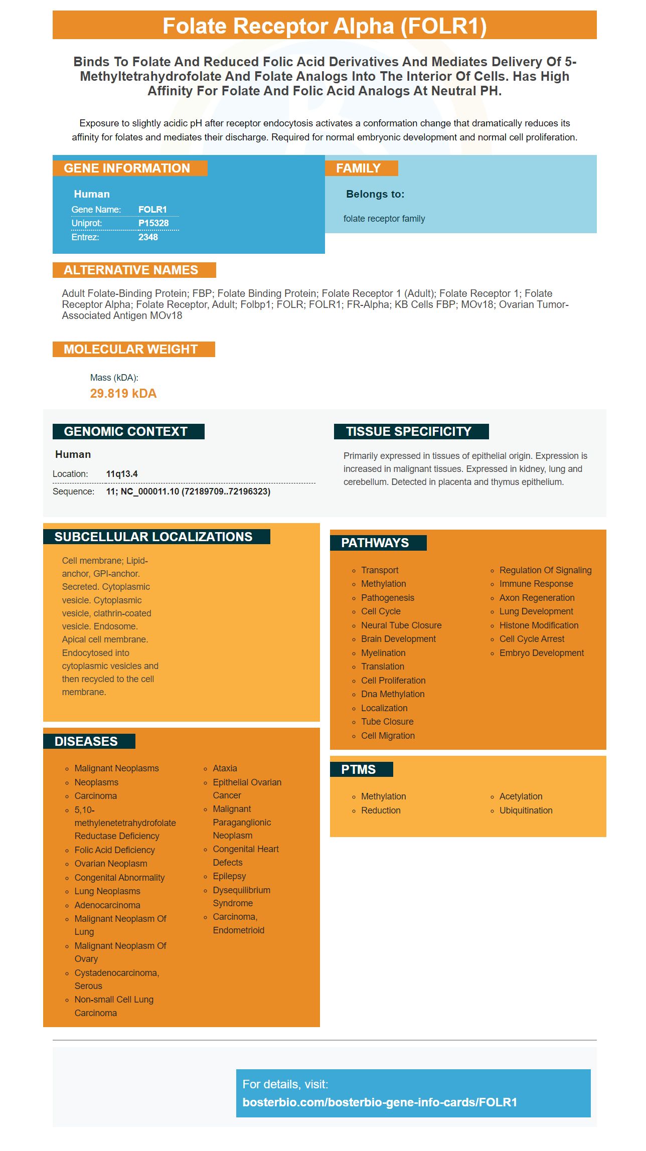
Exposure to slightly acidic pH after receptor endocytosis activates a conformation change that dramatically reduces its affinity for folates and mediates their discharge. Required for normal embryonic development and normal cell proliferation.
Human | |
---|---|
Gene Name: | FOLR1 |
Uniprot: | P15328 |
Entrez: | 2348 |
Belongs to: |
---|
folate receptor family |
Adult folate-binding protein; FBP; folate binding protein; folate receptor 1 (adult); Folate receptor 1; folate receptor alpha; Folate receptor, adult; Folbp1; FOLR; FOLR1; FR-alpha; KB cells FBP; MOv18; Ovarian tumor-associated antigen MOv18
Mass (kDA):
29.819 kDA
Human | |
---|---|
Location: | 11q13.4 |
Sequence: | 11; NC_000011.10 (72189709..72196323) |
Primarily expressed in tissues of epithelial origin. Expression is increased in malignant tissues. Expressed in kidney, lung and cerebellum. Detected in placenta and thymus epithelium.
Cell membrane; Lipid-anchor, GPI-anchor. Secreted. Cytoplasmic vesicle. Cytoplasmic vesicle, clathrin-coated vesicle. Endosome. Apical cell membrane. Endocytosed into cytoplasmic vesicles and then recycled to the cell membrane.
Folate receptors can be used as cancer markers. In this article, we'll learn how the FOLR1 antigen can help researchers find cancer. FOLR1 is an important cancer marker. However, it also serves as a functional proteins. Find out more about the FOLR1 antigen and its uses. Also, read on for some interesting trivia. FOLR1 antigen, a protein found in many tissue types, is the FOLR1 antibody.
The FOLR1 antigen is expressed in many tissues, including the lung. FOLR1 expression levels on alveolar chips are different from FOLR1 expression on HeLa cells, which are commonly used for cancer research. Figure 1E illustrates how the FOLR1 Antigen is expressed throughout the alveoli. This is because cellular growth and folate levels are known to upregulate FOLR1 expression levels. This article will explain the differences between chip and native cells as well as the molecular densities of FOLR1+ chips cells.
FOLR1 has been identified as a marker of many types cancers. Overexpression of the antigen has led to immunotherapy for solid tumors. The antigen is highly sensitive to folate and may initiate intracellular regulatory signals. FOLR1 is also believed to enter and act as transcription factor. FOLR1 knockdowns in ovarian cancer cells inhibit folate-mediated cell proliferation, which prevents their invasive phenotype.
FOLR1–CAR-expressing NK cell symbionts show specificity against folliculin–positive GC cells. FOLR1CAR T cells from human PBMCs were isolated and transduced to the FOLR1CAR lentivirus. Cells were monitored to evaluate the efficiency of lentivirus infection using the CellTiter-Glo assay. After 12 days, the growth rate of infected T cells was not affected by the virus. FACS analysis also examined fluorescences of GFP in the T cell on day 12.
Multiple studies of FOLR1 indicate that this antigen is associated with a prognostic sign for ovarian carcinoma. Its effects on cancer cells are well documented. Treatment of ovarian cancer patients with FOLR1-CAR N cells can help prevent tumors in mice. Further studies are required to confirm FOLR1CAR’s effectiveness in NK cells. The antitumor effect of FOLR1-CAR-NK cells has been shown to be more effective than their counterparts in killing cancer-bearing NOD SCID mice.
Tumor markers can be derived from proteins in tumor cells. These markers are useful in diagnosing cancer and guiding treatment. They may also be useful when combined with other tests to determine a patient's health condition. If you suspect that your cancer may be present, consult your physician immediately. Consider using the FOLR1 Antigen in conjunction with other cancer-related tests.
FOLR1 gene instructs to make a protein called Folate receptor Alpha, which is involved with transporting folate to cells. Folate, also known as vitamin B9, can be used by the body to support many processes. It is absorbed through the intestines and is released as 5-methyltetrahydrofolate, which is taken up by the cells of various tissues.
Folate receptor Beta is a cell type-specific protein and is often found in solid tumors. The expression of this protein is restricted to macrophages, which may represent tumor associated macrophages. As with folate receptor alpha, the expression of FOLR2 is often seen on tumor associated macrophages. This protein is expressed in hematopoietic cells of the macrophage M1 or M2 phenotype.
FRa is a biomarker for the presence of cancer. FRa was previously thought to be a folate transporter. However, it is also a transcription factor. While the relationship between FA and cancer risk remains unknown, studies have shown that FRa can help reduce the risk of many types of cancer. The research team used a mouse-model to study the impact of folate on the growth of ovarian and other cancers. A small study of a manmade molecule called FRa was also done.
Folate plays an essential role in the nucleic-acid synthesis, genome maintenance, amino-acid metabolism, and neurotransmitter/synthetic synthesis. Folate is not synthesized naturally by humans. Instead, we must get the nutrients from our diet. Folate deficiency is a common problem worldwide, and it is associated with a range of diseases. To determine the presence of folate deficiencies, a molecular indicator has been developed.
The folate receptor cDNA is contained in the human genome. Another protein, FRb, encodes the gene. These proteins are involved in folate transport and are anchored to membranes. Folate is highly detectable by these receptors. Endocytosis, which is essential for nutrient absorption, transports folate from the blood into cells.
Folate receptor is a recent evolutionary development. Folate receptor alpha, which was originally called folate receptor alpha in humans, is now called folate b in mice. It is an important marker of the disease as it is a vital gene in mice. It has been linked to many diseases and inherited disorders. Folate is essential for the brain, nervous system and other organs. To prevent folate deficiency, folate levels should be monitored.
Folate has been associated in the prevention of neural tube defects since many years. However, it is now understood to play an important part in adult onset disease. Specific carriers and receptors like FRa can absorb folate. Folate receptor alpha (FRa) is an essential gene for embryonic development, and disruption of both allele leads to severe malformations and lethality at the closure of the neural tube.
Although it is a cancer marker, it is not recommended for all patients. It is helpful in evaluating treatment effectiveness for patients with lung disease. Patients with certain types may also have higher levels of tumor markers, such non-small-cell lung carcinoma. ASCO is also supported by the National Comprehensive Cancer Network (an organization made up of 19 leading US cancer centres). CEA is also an important cancer marker.
CEA is used by many doctors to detect colorectal cancer. CEA levels above five units/mL are generally elevated in liver, thyroid, pancreas and cervix cancer. Additionally, levels of CEA are elevated in healthy people. Elevated CEA levels might indicate more advanced cancer stages. CEA, a cancer marker, can help doctors make a more accurate diagnosis.
Many commercial testing facilities produce tumor markers. Some of these tests do not meet Clinical Laboratory Improvement Amendments guidelines. They may also be considered experimental. Most tumor markers can be used to predict treatment response and prognosis. However, they cannot cure cancer. Consult a doctor if you are interested in any possible treatment. It may also be helpful in screening for cancer patients. You can ask your doctor about the tumor marker results the next time you notice a sign of cancer.
Guidelines for laboratory medicine are published by the National Academy of Clinical Biochemistry. These guidelines address how to use different types or tumor markers. Tumor markers can be used to detect the presence of cancer in blood or urine. They also recommend protocols for biospecimen collection and processing. There are many types of cancer markers available, but not all are suitable for all patients. A cancer marker is useful for patients who are at a specific stage of cancer.
PMID: 2768245 by Elwood P.C.; Molecular cloning and characterization of the human folate-binding protein cDNA from placenta and malignant tissue culture (KB) cells.
PMID: 2527252 by Lacey S.W., et al. Complementary DNA for the folate binding protein correctly predicts anchoring to the membrane by glycosyl-phosphatidylinositol.
*More publications can be found for each product on its corresponding product page