This website uses cookies to ensure you get the best experience on our website.
- Table of Contents
14 Citations 15 Q&As
1 Citations 11 Q&As
3 Citations
2 Citations
Facts about Vascular endothelial growth factor receptor 1.
Can promote endothelial cell proliferation, survival and angiogenesis in adulthood. Its function in promoting cell proliferation seems to be cell-type specific.
Human | |
---|---|
Gene Name: | FLT1 |
Uniprot: | P17948 |
Entrez: | 2321 |
Belongs to: |
---|
protein kinase superfamily |
EC 2.7.10; EC 2.7.10.1; FLT; FLT1; Flt-1; Fms-like tyrosine kinase 1; fms-related tyrosine kinase 1 (vascular endothelial growth factor/vascularpermeability factor receptor); FRT; Tyrosine-protein kinase FRT; Tyrosine-protein kinase receptor FLT; vascular endothelial growth factor receptor 1; Vascular permeability factor receptor; VEGF R1; VEGFR1; VEGFR-1
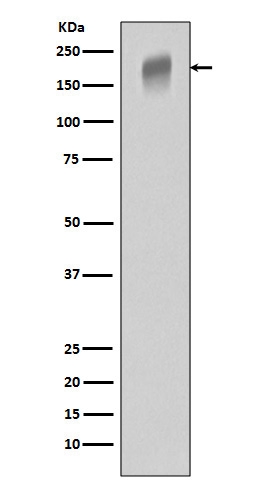
Mass (kDA):
150.769 kDA
Human | |
---|---|
Location: | 13q12.3 |
Sequence: | 13; NC_000013.11 (28300346..28495128, complement) |
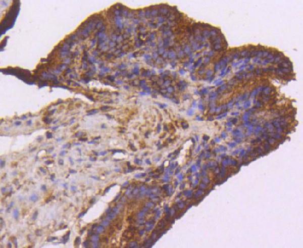
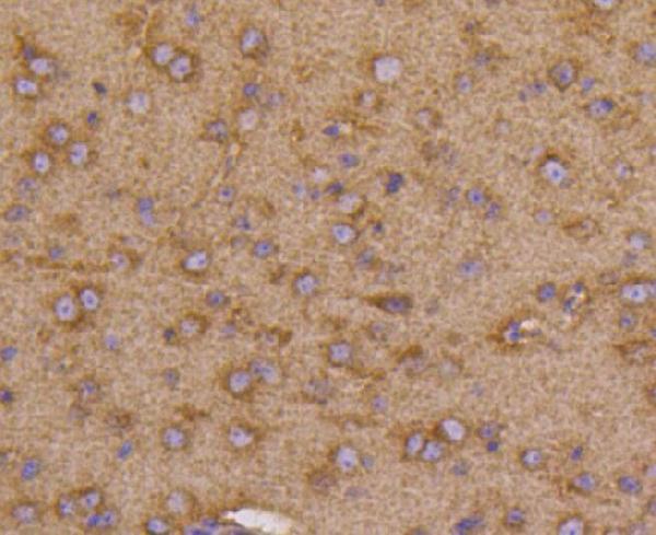
Detected in normal lung, but also in placenta, liver, kidney, heart and brain tissues. Specifically expressed in most of the vascular endothelial cells, and also expressed in peripheral blood monocytes. Isoform 2 is strongly expressed in placenta. Isoform 3 is expressed in corneal epithelial cells (at protein level). Isoform 3 is expressed in vascular smooth muscle cells (VSMC).
[Isoform 1]: Cell membrane; Single-pass type I membrane protein. Endosome. Autophosphorylation promotes ubiquitination and endocytosis.; [Isoform 2]: Secreted.; [Isoform 3]: Secreted.; [Isoform 4]: Secreted.; [Isoform 5]: Cytoplasm.; [Isoform 6]: Cytoplasm.; [Isoform 7]: Cytoplasm.
If you're in the market for an FLT1 marker You've probably heard about Boster Bio. But how do you get the most out of Boster Bio? This article will explore the advantages of the marker, sample preparation and the importance of labeling proteins. In this article, we'll go over some of the most common uses of the FLT1 marker.
The FLT1 marker is a transmembrane receptor found on the cell surface of monocyte-macrophages. This molecule may be useful as a cell surface marker in differentiating between different subpopulations of monocyte-macrophages. This molecule is found in a variety of cell types and could be useful in understanding their function. However it is important to be aware that Flt-1 does not present in all cells. It is only in specific cells that Flt-1 is expressed at the cell surface.
Although Flt-1 does not function as an indicator of prognosis, numerous studies on colorectal cancer have linked it to poor outcomes. These studies have shown that patients suffering from stage II or III colon cancer had lower postoperative survival rates and a higher rate of local cancer recurrences. The FLT1 marker can also be used to assess the extent of cancer-relatedfibrosis.
Researchers found that tumors that had higher levels of FLT1 expression had a higher vascular density in 102 patients. However, this difference was not statistically significant. More studies are needed to determine whether FLT-1 is essential in determining the extent of macrophage-tumor fusion.
Three FLT1 isoforms have been identified in human trophoblast cells which are not detected by the choriocarcinoma cell lines. One is sFLT1-e15a, and the other two are found in the condition of the media. In this study, we examined sFLT1-e15a and sFLT1-i13 in trophoblast cells. To determine if the respective genes are expressed in these tumors, we observed the growth of xenografted mice tumors.
Flt1 is found in trophoblast, mesenchymal and ECs. Flt1-lacZ placentae don't have an fetal endothelial deficiency or a vascular network within the womb. These results indicate that Flt1 expression does not correlate with the structure of the vascular system in the placenta. This marker can be used to identify the presence of FLT1 during pregnancy.
For cell lysates, Dynabeads Protein G were used to perform immunoprecipitation. This antibody is directed against FLT1's first Ig-like domain (KM1730). The proteins that were precipitated were subjected to Western blotting. The purified sFLT1 wasoforms were then concentrated with beads of Heparin-Sepharose. Samples should be frozen at 4°C to allow for analysis of sFLT1 markers.
It is also possible to perform an qPCR-based test to confirm the presence of FLT1 within human cells. The regulation of many tissues and angiogenesis is one of the major functions of FLT1. Numerous Pol II transcription factors are involved in the regulation of FLT1 in both cell types. For instance, DLX5 and SP3 are common in both PE samples, but ZEB2 is lower in one of the two samples.
Polyadenylation site sequences offer better estimates of transcript 3’ ends. The RNA-Seq method has bias towards the 5' end, therefore it is better suited for determining different splicing patterns. For FLT1 expression analysis, polyadenylation-site sequencing yields more precise results for sFlt1 MRNA isoforms. These results can be used to determine the differences in gene expression.
The FLT1 marker is a highly specific and specific fluorescent protein, has a high affinity for both human and mouse cells. Boster Biologicals developed it to recognize proteins of interest in various biological contexts. It is available as a ELISA kit that allows researchers to detect proteins at the picogram level. This antibody is easily accessible through the online store of the company, Tebu-bio.
You'll want to enhance the quality of the sample preparation process if you are doing IHC. Boster Bio has a complete range of secondary antibodies, IHC detection systems, and isotype-specific controls designed specifically for IHC. If you're still unsure of how to optimize your sample preparation for IHC, this guide will provide answers to the most frequently asked questions. Among these are what is the best antibody concentration, what is the best buffer to block and the best way to minimize cross-reactivity.
Immunohistochemistry, also known as IHC, is a technique that detects antigens within cells. This is due to antibodies binding to antigens. BosterBio's IHC guide covers everything from preparation of samples to IHC troubleshooting. The guide contains strategies for overcoming common IHC problems such as high background or weak staining. It also provides a comprehensive overview of available resources for immunohistochemistry. The company has prepared an extensive list of the most important IHC protocols and techniques that can be utilized as a reference.
PMID: 2158038 by Shibuya M., et al. Nucleotide sequence and expression of a novel human receptor-type tyrosine kinase gene (flt) closely related to the fms family.
PMID: 8248162 by Kendall R.L., et al. Inhibition of vascular endothelial cell growth factor activity by an endogenously encoded soluble receptor.
*More publications can be found for each product on its corresponding product page