This website uses cookies to ensure you get the best experience on our website.
- Table of Contents
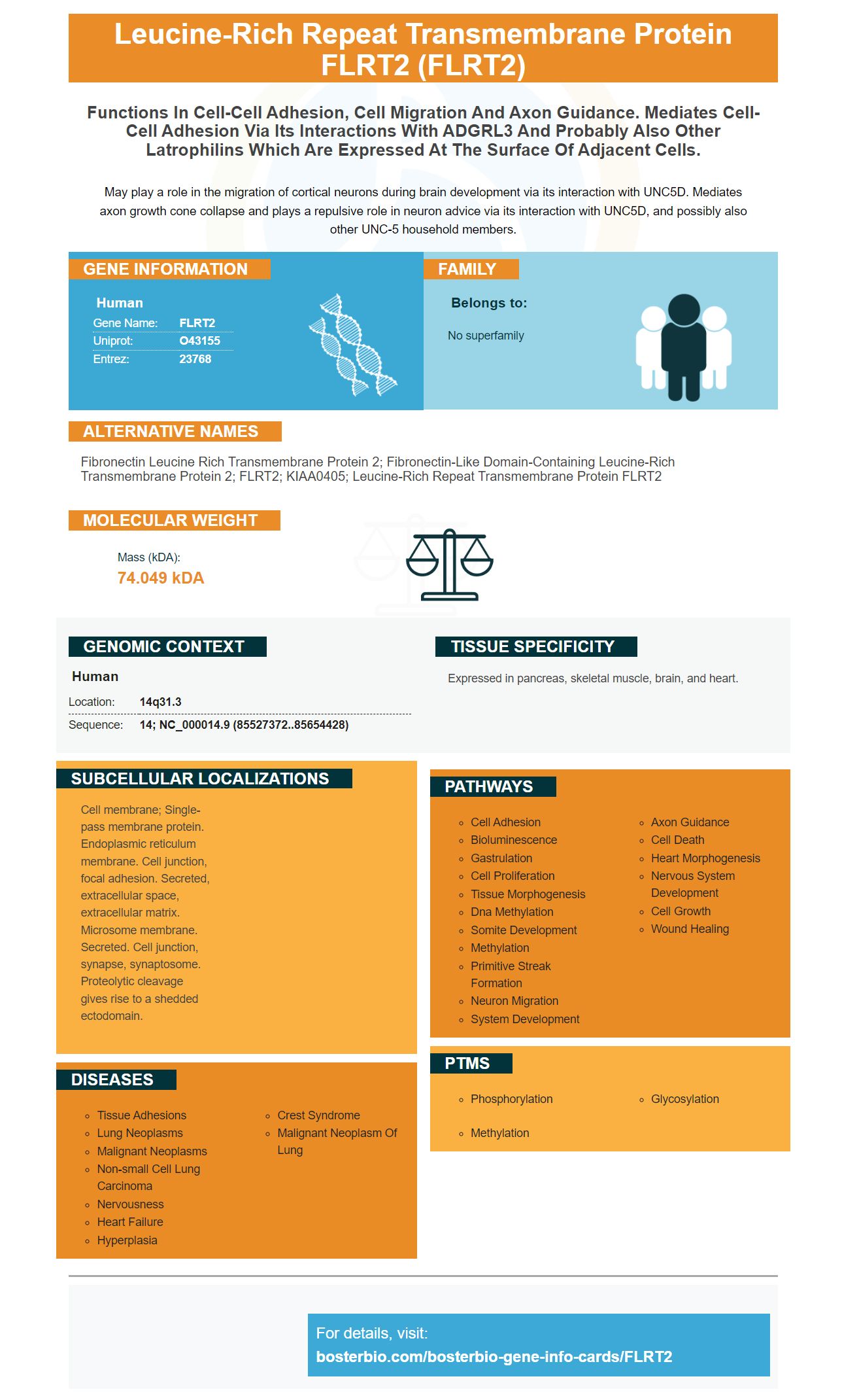
Facts about Leucine-rich repeat transmembrane protein FLRT2.
May play a role in the migration of cortical neurons during brain development via its interaction with UNC5D. Mediates axon growth cone collapse and plays a repulsive role in neuron advice via its interaction with UNC5D, and possibly also other UNC-5 household members.
Human | |
---|---|
Gene Name: | FLRT2 |
Uniprot: | O43155 |
Entrez: | 23768 |
Belongs to: |
---|
No superfamily |
Fibronectin Leucine Rich Transmembrane Protein 2; Fibronectin-Like Domain-Containing Leucine-Rich Transmembrane Protein 2; FLRT2; KIAA0405; Leucine-Rich Repeat Transmembrane Protein FLRT2
Mass (kDA):
74.049 kDA
Human | |
---|---|
Location: | 14q31.3 |
Sequence: | 14; NC_000014.9 (85527372..85654428) |
Expressed in pancreas, skeletal muscle, brain, and heart.
Cell membrane; Single-pass membrane protein. Endoplasmic reticulum membrane. Cell junction, focal adhesion. Secreted, extracellular space, extracellular matrix. Microsome membrane. Secreted. Cell junction, synapse, synaptosome. Proteolytic cleavage gives rise to a shedded ectodomain.
ELISA and flow cytometry are the best methods to measure FLRT2 protein. There are many flow techniques to choose. Boster Bio optimization guide can be used to optimize your experiments. Here are some examples of these guides:
FLRT2 is a common cancer gene, with hypermethylation detected in breast, prostate, lung, and pancreatic cancers. By analyzing gene expression in cancer tissues, cell lines, and other cells, we found that FLRT2 hypermethylation had been detected. In breast cancer cells, FLRT2 levels were both downregulated and increased. These results suggest that FLRT2 could play a role as a tumor suppressor in breast cancer.
FLRT2 regulates many genes, and inhibits cell proliferation. These networks are representative of FLRT2-regulated genes and are grouped by cell type, disease, and functional roles. The top networks of FLRT2 gene expression include immune response to infectious disease, cancer and cell signaling. The heatmap was used to plot gene expression changes. The intensity of the colors represents the magnitude of gene expression changes. Solid and dashed lines indicate the direct and/or indirect interactions of FLRT2 (with other genes)
This method can analyze the entire cell cycle and DNA content in the different phases. Monitoring cell cycle events may help in determining the nature of disease, and provide information to guide treatment and prognosis. Different stages of a cell cycle might reveal altered DNA content or the presence of cancerous cells, as well as the stage at which the cell is dying. The data obtained from this test are stored and analyzed in specialized flow cytometry software. The data are often displayed in the format of a dot chart or histogram.
Flow cytometry is a powerful tool for diagnosing blood and bone marrow cancers. It can detect specific blood cells, such as cancerous lymphoma or leukemia. This test will allow your healthcare provider to provide more effective care based upon the results. However, you must get the permission of your doctor before you undergo flow-cytometry to test for cancer.
Human FLRT2 antibody, labeled using APC-Cy7, detects FLRT2 on a variety of cells. The molecule performs a variety of functions, including cell-cell adhesion and latrophilin-mediated signals as well as axon guidance. The protein is required for normal development and maintenance of the cardiac basement membrane.
Researchers isolated the membrane from giant, unilamellar vesicles that contained the potassium channel KvAP (and the water channel QP0) in order to determine the role membrane curvature plays in cellular function. These membranes contained enriched amounts of KvAP and AQP0, which were indistinguishable in the membranes with higher curvature.
The human FLRT2 ELISA kit from Boster uses sandwich ELISA technology. The monoclonal antibody for human FLRT2 was prepared on 96-well plates. After that, the standards and test samples were added. The unbound conjugates could be removed by washing with PBS. After this, the Avidin-Biotin-Peroxidase complex was added to the plate and incubated for one hour. The FLRT2 quantitative detector kit was used to analyze the protein by comparing it with a standard curve.
Anti-Tja antibody antibodies can be found on endothelial-cell surfaces. This protein can detect human FLRT3. This enzyme-linked assay uses the Double antibody sandwich technique. This test is highly sensitive and has not shown any cross-reactivity to autoantigens from other species.
The proprietary chromatographic methods used to purify human FLRT2 are proprietary. The FLRT2 protein purified can be stored at 4degC up to four weeks or at 20degC longer. The protein should be stored with its carrier protein. It should not stay at room temperature. Information can be modified as needed.
An ELISA for FLRT2 marker is a great way to determine the level of this protein within cell culture. This marker is expressed on many different cell types, including neuronal cells. It has a broad expression throughout the developing nervous system. FLRTs and Unc5s have cell-type-specific expression patterns, but both proteins show promiscuous binding to all other FLRTs. In this way, a cell expressing FLRT2 may interact with any other FLRT-expressing neuron.
FLRT2 is found in many tissues including the craniofacial area. It is found in the vomero–nasal organ, mandibular primarios, and the posterior aspect the secondary palatal shelves. It is also a key component in craniofacial development. FLRT2 is not only expressed in the brain but also in the vomero–nasal organ and mandibular primodia.
A typical ELISA to detect the FLRT2 marker in human serum samples is done. The sample is diluted using 0.05% Tween-20 in a blocking buffer. To achieve desired levels of sensitivity, the blocking buffer can be diluted with detergent. The sample is then incubated for at least 2 hours before the ELISA is read. The results can be interpreted with a slide reader or quantitative immunoblotting.
The FLRT2 gene is expressed in SACs. It also appears in a small fraction of cells in inner nuclear layers (INL). In vitro analysis, FLRT2 expression must colocalize with ChAT+ SAC somas. In vivo FLRT2+ cells must only be seen at the ganglion and inner nuclear layers. However, FLRT2 expression in cell culture experiments is not sufficient to distinguish between SAC and ganglion cells.
In biological assays antibodies against FLRT2 will be used. These antibodies can be monoclonal or multiclonal and react with FLRT2 at different samples. Boster Bio uses antibodies to FLRT2 from rabbit and mouse and focuses on cell-cell adhesion function. It is important for you to know that FLRT2 might also interact with latrophilins.
Many biological applications can be found in the FLRT2 marker. It has been implicated as a cell-cell adhesion marker, as well as in interactions between other latrophilins. There are many antibodies and flow procedures to choose from in your experiment. Boster Bio has compiled a series o optimization tips and guides that will help optimize your experiment. This article will discuss the benefits and drawbacks of FLRT2.
PMID: 10644439 by Lacy S.E., et al. Identification of FLRT1, FLRT2, and FLRT3: a novel family of transmembrane leucine-rich repeat proteins.