This website uses cookies to ensure you get the best experience on our website.
- Table of Contents
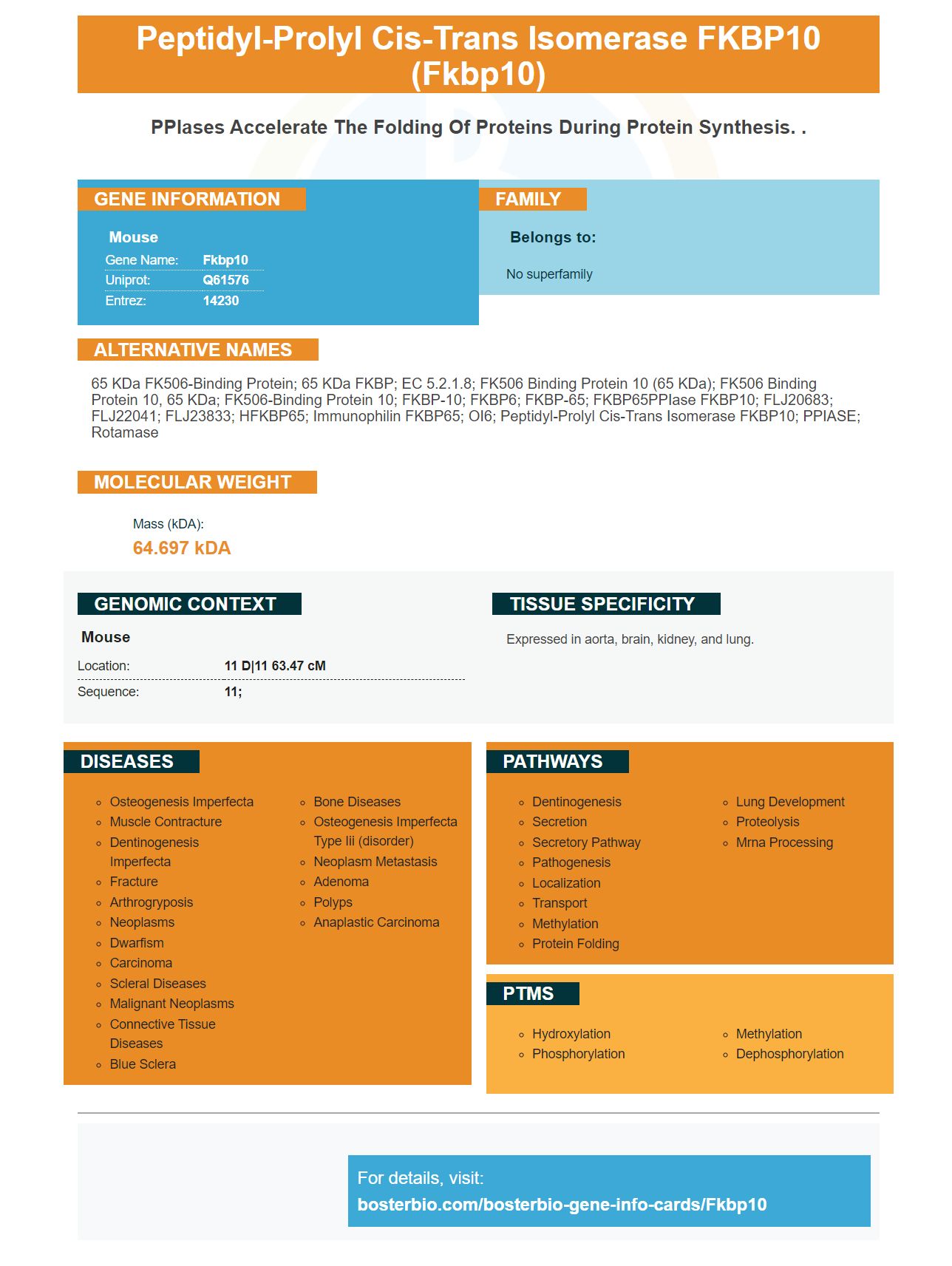


Facts about Peptidyl-prolyl cis-trans isomerase FKBP10.
| Mouse | |
|---|---|
| Gene Name: | Fkbp10 |
| Uniprot: | Q61576 |
| Entrez: | 14230 |

| Belongs to: |
|---|
| No superfamily |

65 kDa FK506-binding protein; 65 kDa FKBP; EC 5.2.1.8; FK506 binding protein 10 (65 kDa); FK506 binding protein 10, 65 kDa; FK506-binding protein 10; FKBP-10; FKBP6; FKBP-65; FKBP65PPIase FKBP10; FLJ20683; FLJ22041; FLJ23833; hFKBP65; Immunophilin FKBP65; OI6; peptidyl-prolyl cis-trans isomerase FKBP10; PPIASE; rotamase
Mass (kDA):
64.697 kDA

| Mouse | |
|---|---|
| Location: | 11 D|11 63.47 cM |
| Sequence: | 11; |
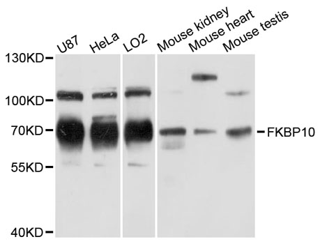
Expressed in aorta, brain, kidney, and lung.


There are many uses of the Anti-FKBP10 Marker. In this article, we will look at detection methods, availability, and proteomic analysis. Lastly, we will look at a case study highlighting the best use of the FKBP10 Marker in the pharmaceutical industry. Read on to learn more. And, don't forget to check out Boster Bio's other great products!
The Anti-FKBP10 Marker in boster bio is an antibody that binds to the FKBP10 gene. FKBP10 is a protein involved in the regulation of cell cycle progression. Its expression is correlated with TNM staging and cancer stage, but not with age, gender, or size of the tumor. It can be used in the diagnosis of CcRCC patients.
This antibody targets the protein FKBP10. It binds to the target proteins in a specific manner. The anti-FKBP10 marker can be used for a number of applications, including cell-based experiments. The Boster Bio Anti-FKBP10 Marker has excellent immunostaining capabilities. This antibody has been validated for use in the clinical setting by the National Institutes of Health.
The FKBP10 gene encodes a protein that binds FK506 and functions as a molecular chaperone. The Boster Bio Anti-FKBP10 Marker uses multiple methods to validate the antibody against a variety of samples that have been tested for a positive result. The FKBP10 gene is found in many tissues, including the brain. Boster's Anti-FKBP10 Marker in Boster Bio is designed to detect the expression of FK506 in multiple tumor types and graded tissues.
The FKBP10 gene interacts with collagen VI, and a deficiency of FKBP10 reduces the migration of phLF. Moreover, FKBP10 is an intracellular regulator of ECM remodeling, and it is a potential drug target for IPF. Its role in preventing the progression of IPF has been demonstrated by multiple studies. So, the Anti-FKBP10 Marker in Boster Bio is highly regarded.
To detect mutations in the FKBP10 marker, Sanger sequencing has been used. The study analyzed samples from 100 unrelated and ethnically matched individuals. The result of this test is a copy number variation in the gene. The mutations were confirmed by comparing the obtained sequences with a reference sequence of the gene. This analysis is a promising method for the early detection of rare and hereditary diseases.
The detection of the FKBP10 marker can also be performed using a Western blot. The markers analyzed include integrin a1, a2, aV, and a6. The analysis was performed using b-actin as a loading control. Western blots show the protein expression of these proteins. For example, the integrin a1 protein shows the greatest degree of expression in the bone-marrow of a patient.
This analysis also reveals that FKBP10 has an important role in adhesion in GC cells. It was also found that silencing the FKBP10 gene in GC cells reduced cell adhesion. As FKBP10 is a GC cell-specific biomarker, this protein may help in the treatment of this disease. The study has also found that it is a viable biomarker to monitor lymph node metastasis.
Marker gene selection is a complex process. Expert knowledge, statistical tests, and mean-variance optimization were used to identify the best marker candidates. These methods could be automated using supervised learning approaches. The process of selecting markers is a repeatable one as the database grows. Detection methods of the FKBP10 marker
In recent years, proteomic analysis has made tremendous progress. Advances in proteomics technologies have made it possible to identify protein species from diverse sources, while improving detection sensitivity and achieving broader proteome coverage. Increased application of proteomics techniques has benefited research across fields related to biology, such as structural and sequence analysis. However, further improvements in these technologies are required to improve the reproducibility and performance of well-known tools.
High-throughput methods have made bioinformatics a vital component of proteomics. In particular, these methods depend on robust data analysis. Bioinformatics provides novel algorithms for proteomics data management and advances the discovery process. This section provides an overview of some of the most common approaches used in proteomic analysis. To get started, consider the following workflows. Typical experimental workflows include label-free quantification and shotgun proteomics.
Sample preparation. The preparation of plant materials depends on the species, stage of development, and protein fragment being analyzed. Depending on the sample material, each plant cell type can contain a specific protein population. Therefore, it is important to select the correct extraction protocol for your research objectives. Further, there are several protein extraction methods, each with its own set of pros and cons. The choice of one should depend on the type of plant material and your desired results.
Plants and pathogens. Plant tissues are difficult to disrupt, and they contain a high level of secondary metabolites that can interfere with the analysis. Pathogens are also difficult to prepare for proteomic analysis. They are difficult to cultivate on artificial media and are long-lived, resting spores. The following examples demonstrate the practical application of proteomics for plant biology. This application is increasingly gaining ground, but still requires some innovation.
The expression of FKBP10 in cancer cells and tissues has been associated with poor prognosis. This gene is involved in cell adhesion, proliferation, and apoptosis, and low expression in CcRCC may indicate poor prognosis. The authors declare that they have no competing interests. These data were obtained using the GEPIA web server, which allows users to perform gene expression profiling and interactive analysis. The data are licensed under the Creative Commons Attribution License.
The FKBP10 gene encodes a protein with four PPIase domains and a CA-BIND domain. One of the variants in the gene encodes a nonsense codon in the EF-hand domain. The deletion of this codon likely impaired the protein's function and reduced its size. It may also affect the mineralization of the matrix. These data may suggest the role of FKBP10 in skeletal development.
Collagen VI is the most important part of the extracellular matrix (ECM). The protein is essential for the formation of collagen and elastin. Without collagen, it is degraded by proteolytic enzymes and may not contribute to the higher ordered organization of the ECM. Therefore, downregulation of FKBP10 may result in disorganization of the ECM and decreased crosslinking sites in type I and type VI collagen.
The availability of FKBP10 in the lung has been reported in two independent cohorts of patients with IPF. These cohorts showed that FKBP10 expression was found in fibrotic mouse lung tissue as well as primary human lung fibroblasts. Both fibrotic lung tissues and cell adhesion and migration were impaired when FKBP10 was deficient in either tissue. These results show that FKBP10 is an important fibrotic gene in IPF.
The FKBP10 marker is a protein that is found in the endoplasmic reticulum of the kidney. This organelle is involved in protein processing, transport, and production. It is vital for the correct processing of collagen, a protein that gives strength to connective tissues. The cost of the marker depends on how often you want to test it. There are several tests available. To know how much it will cost, see the pricing table below.
A recent study showed that FKBP10 significantly decreased integrin a6 and aV in the cytoskeleton of GC cells. This suggests that FKBP10 may act through the integrin/AKT pathway to promote cell adhesion and migration. However, a number of researchers have questioned whether or not FKBP10 is involved in fibroblast migration. They believe that FKBP10 is required for the fusion of collagen and elastin.
The FKBP10 gene is associated with cancer. Recent research has shown that it is negatively correlated with survival in gastric and lung cancer patients. However, the exact mechanism is not yet clear. However, it is known that FKBP10 affects cell cycle progression and regulates invasion of the ccRCC cell line. Further research is needed to understand the FKBP10 gene's function in cancer.
As an intracellular receptor for the immunosuppressant FK506, FKBP10 plays an important role in tumor development. It has also been shown to regulate the PI3K signaling pathway, and is also associated with prostate cancer carcinogenesis. FKBP10 remains an important molecular mechanism for the progression of clear cell renal cell carcinoma. The cost of the FKBP10 gene marker will depend on the amount of cancer you have.
PMID: 7507077 by Simek S.L., et al. Sequence and localization of a novel FK506-binding protein to mouse chromosome 11.
PMID: 7493967 by Coss M.C., et al. Molecular cloning, DNA sequence analysis, and biochemical characterization of a novel 65-kDa FK506-binding protein (FKBP65).