This website uses cookies to ensure you get the best experience on our website.
- Table of Contents
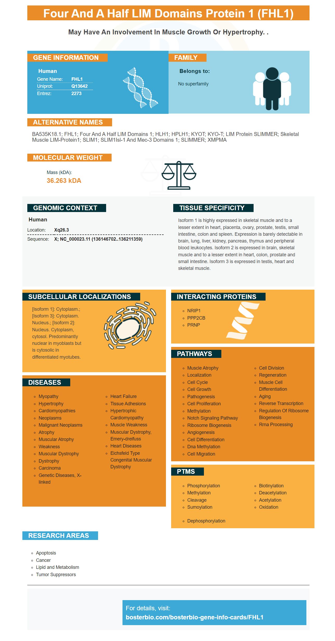
Facts about Four and a half LIM domains protein 1.
Human | |
---|---|
Gene Name: | FHL1 |
Uniprot: | Q13642 |
Entrez: | 2273 |
Belongs to: |
---|
No superfamily |
bA535K18.1; FHL1; four and a half LIM domains 1; HLH1; HPLH1; KYOT; KYO-T; LIM protein SLIMMER; skeletal muscle LIM-protein1; SLIM1; SLIM1Isl-1 and Mec-3 domains 1; SLIMMER; XMPMA
Mass (kDA):
36.263 kDA
Human | |
---|---|
Location: | Xq26.3 |
Sequence: | X; NC_000023.11 (136146702..136211359) |
Isoform 1 is highly expressed in skeletal muscle and to a lesser extent in heart, placenta, ovary, prostate, testis, small intestine, colon and spleen. Expression is barely detectable in brain, lung, liver, kidney, pancreas, thymus and peripheral blood leukocytes. Isoform 2 is expressed in brain, skeletal muscle and to a lesser extent in heart, colon, prostate and small intestine. Isoform 3 is expressed in testis, heart and skeletal muscle.
[Isoform 1]: Cytoplasm.; [Isoform 3]: Cytoplasm. Nucleus.; [Isoform 2]: Nucleus. Cytoplasm, cytosol. Predominantly nuclear in myoblasts but is cytosolic in differentiated myotubes.
The FHL1 marker could be a new diagnostic or prognostic tool to help patients suffering from glioma. It could also function as an anti-tumor agent and a promising therapeutic target in future. In this article, we'll look at the FHL1 marker and its potential applications. The information is provided by Sanbio. We also discuss the limitations and benefits of the FHL1 marker and what it means for the future of glioma therapy.
A cytotoxic enzyme, the granzyme B activates caspases and other DNA repair factors in the Apoptotic pathway. This causes the release of cytochrome c and the activation of caspases-3 6, and 7. Both enzymes cause death of target cells by the cleavage process of DFFA or bid. Granzyme B also removes Mcl-1, an anti-apoptotic protein. It triggers the release of the Bcl-2 family member, pro-apoptotic Bim, which further damages mitochondria.
The crystal structure of granzyme shows the specificity of the substrate for the enzyme. In the structure, the two molecules that are bonded to ecotin are shown. The orange atoms are key residues of catalytic sites. A diagram of enzyme's 2.2A electron densities show that the primary determinant for substrate specificity is residue Arg226. Three hydrogen bonds between Gln217, Ser221, and Ser226 keep the Arg226 residue in place. The substrate aspartic acid is illustrated in blue.
The human granzymeB gene measures around 3.2 km long. This gene is considered a prototype for granzyme gene organization. The first exon code a leader sequence, while the second exon codes individual amino acids of a catalytic triad. Exons II and I correspond to the granzyme A and the gene cathepsin G on the chromosome 19p13.3.
A recently discovered tumor-specific fusion gene, the EGFRvIII mutation, displays radiomic evidence of the glioma. This mutation has been associated with higher rCBVlevels, lower ADC and a decrease in T2-FLAIR. Furthermore, tumors carrying the EGFRvIII mutation are often found to overlap with the temporal and frontoparietal lobes.
The genetics of gliomas were determined by using two subgroups of the current study. The first group is located along the astrocytic path and has mutations in ALT, TP53 and IDH. The second group has a distinct histologic profile. It is affected by mutations in CIC and FUBP1, FGFR, TERT promor, FUBP1, 1p/19q, and CIC. The second group has a higher median overall survival.
At present, histological grade is the most reliable marker for diagnosing and prognosis for a patient's outcome in clinical. The majority of tumors with higher gliomas exhibit faster uptake and washout. PET imaging with FDOPA has 82% accuracy in determining true and pseudo-progression in the glioblastoma. However, there is a challenge in determining whether a patient has 1p19q codeletion or not.
Patients with high-grade gliomas had higher levels of plasma small-sized DNA than those who had low-grade gliomas. Patients with low-grade glioma also had higher levels of small-sized DNA in their blood. It is not clear how IDh2 mutations differ from those in IDH2, but the two genes have the same impact.
Gliomas are more vulnerable to immunotherapy because of a common mutation. This could have broader therapeutic implications. The findings of a new mouse model showed that a single mutation in the tumor may hold the key to more efficient immunotherapy. Researchers are hoping to develop treatments for immunotherapy that target this single gene in the near future. Here are some potential therapeutic benefits for FHL1.
A mouse model of glioma was discovered to show that FHL1 delivery through adenovirus decreased tumor growth by cutting down VEGF expression. The same method also inhibited the growth of angiogenic tumors in mice. Furthermore, the delivery of intratumorally administered antisense RNA against the TK-domain slowed the growth of gliomas.
While the p53 gene has been identified as an glioma-related target but it isn't known what causes this cancer gene to be often inactivated. In the glioma process, this gene regulates many different cellular functions such as cell-cycle regulation, cellular growth, apoptosis, as well as DNA damage repair. Therefore targeting FHL1 for research on glioma is an exciting direction to pursue in the near future.
A new study has demonstrated that the reduction of MGMT genes in glioma cells could increase their sensitivity to TMZ. Additionally, a new liposome that targets FHL1 was designed. In addition two peptides T7 and A7R improve the transfer of plasmid DNA across the BBB. Both PBAE-based nanoparticles aswell with HSV-TK DNA loaded liposomes were successful in targeting brain glioma cell lines the test tube.
FHL1 is a tumor suppressor. It blocks anchorage-dependent growth of human hepatocellular cancer cells. FHL1 interacts with SMAD proteins in a non-dependent manner with TGF-b which promotes nuclear translocation of Smad4 that blocks the oncogene c-myc and tumor suppressor gene p21. FHL1's inhibition of the hepatocellular carcinoma cells require CK1d.
To study the role of FHL1 in NSCLC we utilized miR-4502 as a tumor suppressor. MiR-4502 blocks the expression of FHL1 by binding to the 3 untranslated 3' region. It is a new target for miR105 and blocks FHL1. It hinders NSCLC cells' metastasis, proliferation and viability by increasing FHL1.
The four and a half LIM domains of FHL1 interact with different molecules including DNA and RNA. FHL1's zinc-finger domain regulates signal transduction. It also plays a role in gene transcription, cytoarchitecture and transcription of genes. FHL1 has been linked with myopathies of various kinds. However, the protein is not yet fully understood. In addition to being a tumor suppressor FHL1 is also a neurodegenerative proteins.
A molecular analysis of the expression of FHL1 in human gastric cancer cell lines reveals an association between decreased FHL1 expression and aggressiveness. Patients with primary gastric cancer will have a lower survival rate in cases where FHL1 is decreased. Overexpression of FHL1 is associated with a lower rate of disease progression and a shorter time to death. FHL1 is an anti-tumor agent and is a key factor in the development of cancer.
Several pathways are activated when FHL1 is expressed. The TGF–b1/Smad and Wnt/b–catenin pathways are two of them. The activation of FHL2 increases B-catenin signalling, and the opposite effect is inhibited by down-regulation. It is interesting to note that FHL2 has a role in tubular EMT.
TGF-b1 is an agonist which induces the expression of FHL2 mRNA in the NRK-52E cells. It also stimulates the expression of FHL2 protein in a time and dose-dependent way. Additionally, FHL3 knockdown inhibits the growth of gastric cancer cells in a way that reduces their metastasis as well as facilitating treatment.
The PTEN/PI3K/Akt pathway regulates cell proliferation, differentiation, and autophagy. This protein is a lipidphosphatase hydrolyzing the phosphates in phosphoinositides. In the mdx muscle, FHL1 expression contributes to the inhibition of the PI3K/AKT pathway. Muscle cells in patients with muscular dystrophy express FHL1 markers, as are Duchenne muscular dystrophy sufferers.
FHL1 regulates PI3K/AKT signals and also regulates expression of mTOR, Bcl-2. The expression of these proteins also regulates apoptosis within leukemia cells. These results indicate that the FHL1 marker is responsible for the PI3K/AKT signal in a manner similar as its role in tumorigenesis.
For miR410-mediated proliferation of M cells for MM cells to multiply, the FHL1 gene must be present. It also inhibits cell cycle progression and the process of apoptosis. In addition, miR-410 promotes cell proliferation in MM cells through targeting KLF10.
The FHL1 marker is a component of the PI3K/AKT pathway can be used to pinpoint potential drugs for the treatment of cancer. It hinders the growth of various types of cancer cells through interfering with the activity of AKT/PI3K. FHL1 expression also reduces the growth of different types of gliomas.
As commercially accessible products anti-FLL1 antigens are available on the market. Boster Bio develops antibody kits that target biomarkers in various fields, including neuroscience cancer, and the development of biology. The effectiveness of these antibodies can be up to picogram levels. Boster Bio sells its products through tebu bio. Boster Bio also produces research antibodies and immunological reagents for the study of biomarkers in cancer cells.
PMID: 8753811 by Morgan M.J., et al. Slim defines a novel family of LIM-proteins expressed in skeletal muscle.
PMID: 9714789 by Lee S.M.Y., et al. Chromosomal mapping, tissue distribution and cDNA sequence of four- and-a-half LIM domain protein 1 (FHL1).