This website uses cookies to ensure you get the best experience on our website.
- Table of Contents
2 Citations 1 Q&As
Facts about Bis(5'-adenosyl)-triphosphatase.
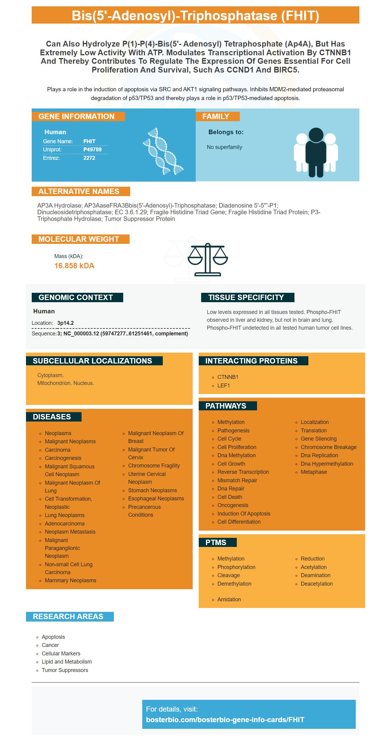
Plays a role in the induction of apoptosis via SRC and AKT1 signaling pathways. Inhibits MDM2-mediated proteasomal degradation of p53/TP53 and thereby plays a role in p53/TP53-mediated apoptosis.
Human | |
---|---|
Gene Name: | FHIT |
Uniprot: | P49789 |
Entrez: | 2272 |
Belongs to: |
---|
No superfamily |
AP3A hydrolase; AP3AaseFRA3Bbis(5'-adenosyl)-triphosphatase; Diadenosine 5'-5'''-P1; dinucleosidetriphosphatase; EC 3.6.1.29; fragile histidine triad gene; Fragile histidine triad protein; P3-triphosphate hydrolase; tumor suppressor protein
Mass (kDA):
16.858 kDA
Human | |
---|---|
Location: | 3p14.2 |
Sequence: | 3; NC_000003.12 (59747277..61251461, complement) |
Low levels expressed in all tissues tested. Phospho-FHIT observed in liver and kidney, but not in brain and lung. Phospho-FHIT undetected in all tested human tumor cell lines.
Cytoplasm. Mitochondrion. Nucleus.
Are you thinking about using the Boster Bio PLG Marker? If yes, you've come to the right spot. This article will provide information on Boster Bio's CSF GM antibody and the PLG Marker. We will also talk about the advantages of using these products. You will be able to use them as needed. We hope that you enjoy this article!
Boster Bio's PLG Marker, biomaterial recently developed can be used to target bacterial antibodies, GM CSF, or CpG. The PLG marker is an effective inhibitor of angiogenesis and could have a number of useful applications in the field. Detailed research will be required to determine the most effective use for this biomaterial. This tool is available in a variety of sizes and shapes.
Boster Bio PLG antibody reacts to human. The Boster Bio Anti-Angiostatin K1-3/PLG antibody is the catalog number RP1027 and is stored at -20°C for up to one year. It is also stable at 4degC for a month. The Boster Anti-Angiostatin K1-3 antibody is a recombinant protein derived from E. coli.
The GM-CSF antibody, produced by Boster Bio, utilizes the FHIT marker. This antibody has been proven in mice to reduce the Abeta1-42 activity as well as microglia within Tg2576. This could have therapeutic implications for Alzheimer's. Further research is required to confirm the anti-inflammatory properties of the antibody. As with all antibodies Boster Bio has patented the FHIT marker.
The GM-CSF antibody is built on the FHIT marker. This molecule is expressed in bone marrow cells. It is the most specific antibody available today. It recognizes both the GM-CSF of animals and humans. It has a standard structure so that you don't need any worries about contamination. Boster Bio GM CSF antibody also uses the FHIT marker, which guarantees it can recognize all kinds of cells.
The ILC3 contains the IL-23R/GMCSF axis. This region of the intestinal wall is essential for regulating the spatiotemporal dynamism of the ILCs. It is also involved in the accumulation of innate effector cells. Additionally, it enhances monocyte viability through GM-CSF. Boster Bio's GM CSF antibody utilizes FHIT to detect intestinal inflammation.
CSL311 by Boster Bio is yet another anti-GM CSF antibody. It targets the GM–CSF-Rb molecule which is similar to IL-3 or IL-5. It is currently being evaluated in phase I to determine if the medication is safe for use in asthma patients. The results of this trial will be made available within the first quarter of 2014.
The use of the FHIT Marker is beneficial in various types of cancer. It can be used to determine patients who may benefit from adjuvant therapy. For instance, the loss of FHIT expression in a patient suffering from non-small cell lung cancer is related to a poor clinical outcome. It can also aid in determining whether or not the cancer will respond to chemotherapy.
PMID: 8598045 by Ohta M., et al. The FHIT gene, spanning the chromosome 3p14.2 fragile site and renal carcinoma-associated t(3;8) breakpoint, is abnormal in digestive tract cancers.
PMID: 9012482 by Druck T., et al. Structure and expression of the human FHIT gene in normal and tumor cells.
*More publications can be found for each product on its corresponding product page