This website uses cookies to ensure you get the best experience on our website.
- Table of Contents
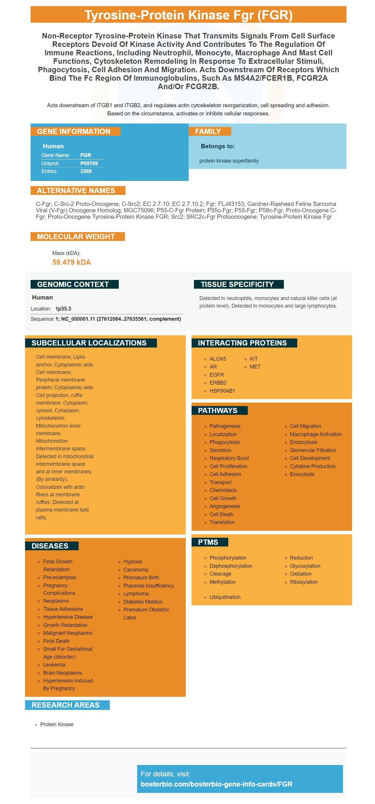
Facts about Tyrosine-protein kinase Fgr.
Acts downstream of ITGB1 and ITGB2, and regulates actin cytoskeleton reorganization, cell spreading and adhesion. Based on the circumstance, activates or inhibits cellular responses.
Human | |
---|---|
Gene Name: | FGR |
Uniprot: | P09769 |
Entrez: | 2268 |
Belongs to: |
---|
protein kinase superfamily |
c-fgr; c-src-2 proto-oncogene; c-src2; EC 2.7.10; EC 2.7.10.2; Fgr; FLJ43153; Gardner-Rasheed feline sarcoma viral (v-fgr) oncogene homolog; MGC75096; p55-c-fgr protein; p55c-fgr; p55-Fgr; p58c-fgr; Proto-oncogene c-Fgr; proto-oncogene tyrosine-protein kinase FGR; Src2; SRC2c-fgr protooncogene; tyrosine-protein kinase Fgr
Mass (kDA):
59.479 kDA
Human | |
---|---|
Location: | 1p35.3 |
Sequence: | 1; NC_000001.11 (27612064..27635561, complement) |
Detected in neutrophils, monocytes and natural killer cells (at protein level). Detected in monocytes and large lymphocytes.
Cell membrane; Lipid-anchor; Cytoplasmic side. Cell membrane; Peripheral membrane protein; Cytoplasmic side. Cell projection, ruffle membrane. Cytoplasm, cytosol. Cytoplasm, cytoskeleton. Mitochondrion inner membrane. Mitochondrion intermembrane space. Detected in mitochondrial intermembrane space and at inner membranes (By similarity). Colocalizes with actin fibers at membrane ruffles. Detected at plasma membrane lipid rafts.
Boster has the best FGR Marker antibody. With its line of high-affinity primary antibodies, Boster provides antibodies that are cited in the research community and validated on Western Blotting, Immunohistochemistry, and ELISA. Here is a closer look at Boster FGR markers. Boster also has high-affinity secondary antibodies.
This book will help you improve your shooting by providing an FGR marker review. Steven Boster owns AB-PT. Inc. and a business-studio with over ten decades of experience. He is a well-respected expert in the field and writes from his own experience. You'll find many ways to use his book, which is full of useful tips and tricks for FGR markers.
Boster Bio is the right company to contact if you are interested in high-affinity prima antibodies for your research. The company specializes on manufacturing picogram sensitive ELISA kits as well as IHC-optimized, polyclonal antibodies. The company has over 12,000 different antibodies that are all validated for IHC, WB, and Flow applications. These antibodies are also based on proprietary trade secrets.
These antibodies are specific for the target antigen and bind only to it, as their name implies. These antibodies are generally classified according to their specificity and affinity. The higher their affinity, the less likely they are to bind to unintended antigens. However, antibodies with high quality have the ability to detect and purify antigens. They are therefore the best choice for research studies.
These monoclonal antibodies are highly specific for the SARS-2 antigen and are therefore useful in biomedical applications. These antibodies can also disrupt high-affinity interactions between the SARS/CoV-2 RBD (human ACE) and these antibodies. High-affinity antibodies were identified using memory cells derived form immunized mice. To carry out single cell screening, sufficient amounts of IgG1 cells were isolated.
A series ELISA testing is necessary to establish the presence of primary antibodies. A titration ELISA test is performed for each test-bleed following immunization to identify supernatants. Positive supernatants are then subjected to limiting-dilution subcloning until the supernatant growth in all wells is similar. Only then are the antibodies selected to be used in further testing.
The flow cytometry was used to characterize the isolated antibodies after purification. They were then compared with biotinylated SARS/CoV-2RBD immobilized in magnetic beads. The EC50 value of the lead clones (12H2 &13I1) and a control antibody (CB6) was determined. All results were based upon three independent experiments. They had high affinity against SARS CoV-2 RBD.
In a sandwich ELISA test, the detection and capture antibodies must recognize different non-overlapping epitopes. Their dynamic range, sensitivity, and dynamic range may be limited when the detection and catch antibodies are the identical. Boster Bio produces high-affinity primary antibodies that are designed for sandwich ELISA. When creating sandwich ELISAs, you should use matched pairs.
During the purification process, proteins containing Protein A were separated from the media. These beads were then rinsed in 75-150mLs of PBS and eluted from the buffer with 0.1M glycine. They were then treated with 1M tris to pH 7.4 in order to reduce their potential for aggregation. The final products were stored at 80 degrees C and checked for purity using SDS-PAGE.
The ELISA technique can be used to measure FGR proteins in cell culture supernatants. You will need high affinity antibodies and a 0.2 um filter. Follow these instructions to get the best results. After you've obtained the results, you can share the results. Boster Bio offers custom services.
Picokine(tm), a proprietary ELISA platform enables high sensitivity down to the picogram level. Its ELISA kit has been tested with a wide range of samples. Images and validation methods are available upon request. Supervision, a secondary antibody based on polymers, saves you 30 minutes of IHC. Boster Bio's technology is supported by knowledge about the design of immunogens. Their customer support team is unmatched.
Cells were grown in complete medium for 48-hours to test FGR-related cells activities. After 48 hours, cells were harvested and processed for enzyme-linked immunosorbent test (ELISA), western blotting or Transwell. In this method, cells were cultured for wound healing, tube formation, and cell proliferation. When the cells were harvested, they were measured for apoptosis as well as proliferation.
PMID: 3275868 by Katamine S., et al. Primary structure of the human fgr proto-oncogene product p55c-fgr.
PMID: 2852026 by Brickell P.M., et al. Structure and expression of c-fgr protooncogene mRNA in Epstein-Barr virus converted cell lines.