This website uses cookies to ensure you get the best experience on our website.
- Table of Contents
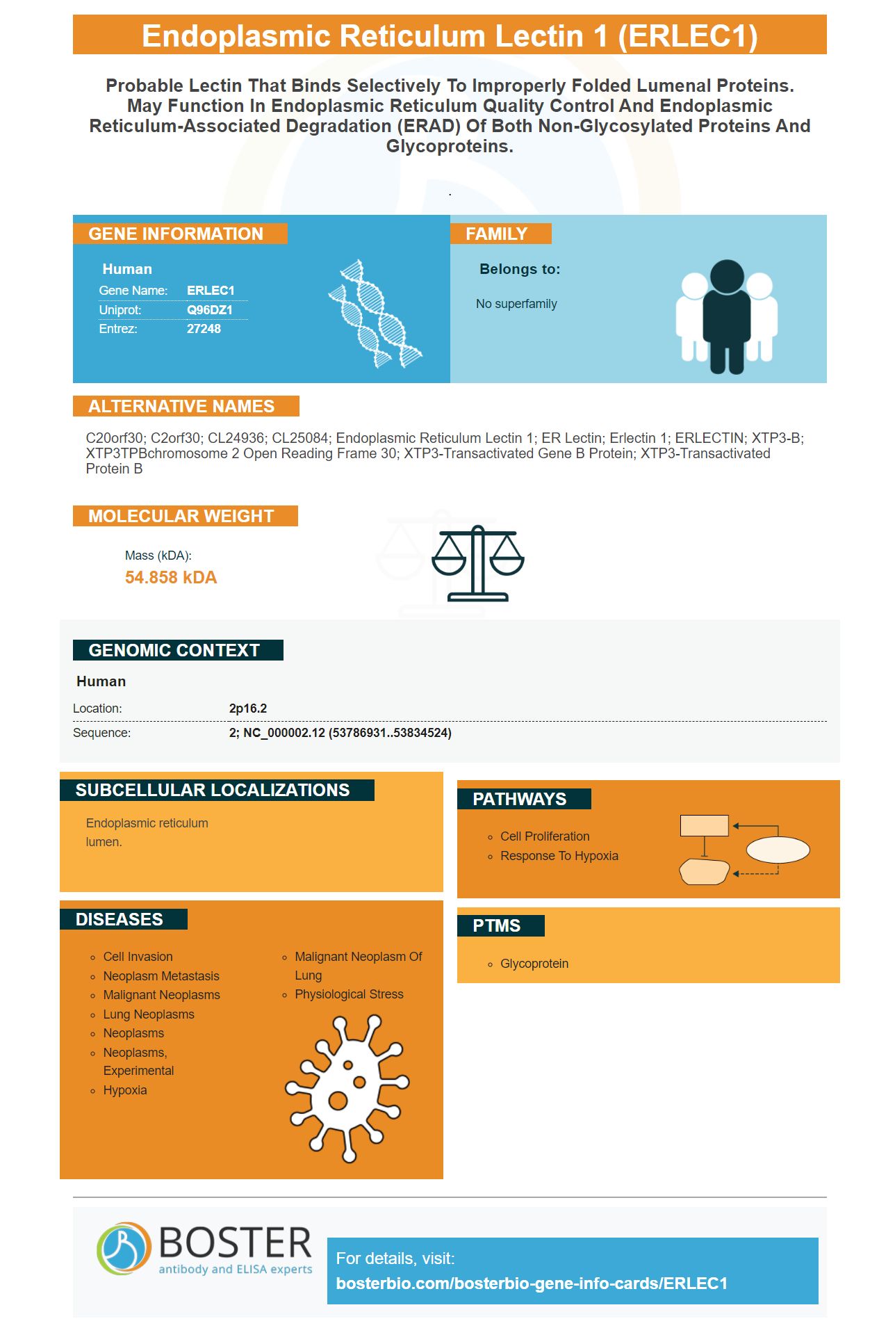
Facts about Endoplasmic reticulum lectin 1.
.
Human | |
---|---|
Gene Name: | ERLEC1 |
Uniprot: | Q96DZ1 |
Entrez: | 27248 |
Belongs to: |
---|
No superfamily |
C20orf30; C2orf30; CL24936; CL25084; endoplasmic reticulum lectin 1; ER lectin; erlectin 1; ERLECTIN; XTP3-B; XTP3TPBchromosome 2 open reading frame 30; XTP3-transactivated gene B protein; XTP3-transactivated protein B
Mass (kDA):
54.858 kDA
Human | |
---|---|
Location: | 2p16.2 |
Sequence: | 2; NC_000002.12 (53786931..53834524) |
Endoplasmic reticulum lumen.
For years, scientists have been wondering how to use the Endoplasmic reticulum lectin 1 (ERLEC1) marker. This article will provide an overview of ERLEC1's specificity, function, and applications. We will be covering the most popular uses of ERLEC1 and how you can submit results to species or applications. These methods are available to all scientists worldwide.
The endoplasmic reticulum contains approximately one-third the human genome. It encodes proteins that are involved with cell surface, secretion, organelle function, and organelle function. When proteins are synthesized, they are translocated to the lumen of the ER where they undergo a series of modifications and fold cotranslationally. Resident molecular Chaperones monitor this process and prevent the proteins aggregating or folding off-pathway. These quality-control steps ensure that only molecular protein forms pass through ER.
This marker was originally derived directly from the DDPCR fragment. The sequence contains the polyadenylation molecular motif (underlined with an angled dash line), the amino compounds comprising N-linked glycosylation locations, and the transmembrane. The mMCL sequence combines the amino acids of the first exon. Future detection of cancer cells will be possible using the intron/exon splice (arrowheads).
ERLEC1 is an endoplasmic reticulum protein. It is involved ER-associated destruction, N-glycan detection, and membrane-associated ubiquitin ligase. It regulates multiple cellular stress response pathways and promotes metastatic cancer survival. Alternative splicing allows for multiple transcript variants of this marker. A pseudogene that is related has been identified for chromosome 21.
Reference markers have been created by the Human Protein Atlas project for different organelles. The Organelle Marker Panel targets 11 subcellular structure in the cell. One of these markers is ERLEC1. When used in WB and IHC applications, it is recommended to use the antibody against calreticulin as a reference. This marker can be used to visualize subcellular distribution patterns. This marker was selected by the Human Protein Atlas project to be a reference for ER.
Based on ERLEC1 expression in breast cancer cells, the study identified candidate markers to detect brain metastasis. These markers may be able to distinguish ErbB-2+ cancers from ErbB2-negative. The ERLEC1 gene can also be used as a biomarker for a variety of breast cancer types. These markers can be used to improve patient care. More research is needed.
In addition to ERLEC1, the markers GRP94, FN14, and TRAF2 were also associated with brain metastasis in the study. Multivariate logistic analysis showed that ERLEC1 & FN14 were associated better with brain metastasis detection than ErbB-2. Combining these markers however gave the best accuracy. The results of this study indicate that the four markers are associated with the brain metastasis in a more complex way than their individual components.
The epigenetic regulator of cancer cell proliferation is the ERLEC1 gene. The gene is expressed in different types of cancers and has been implicated with brain metastases. To further examine the marker, researchers conducted gene expression analysis using samples of normal and breast cancer patients. A positive marker expressed as ERLC1 indicates that a patient’s cancer cell growth is restricted in the peritoneum. While a negative marker expressed only by the glial, there is no clear indication.
The ERLEC1 gene, which is expressed maternally, increases during Xenopus differentiation. This marker is expressed at a higher level during neuronulation. However, this gene has yet to be fully characterized in flies. Scientists must first confirm that ER stress is induced via RNAi-depletion. Other methods to test for ER stress include the Xbp1 transcripts or the Cnx99a antibodies.
In a mouse embryograft model, ERLEC1 deficiency was also associated a poor prognosis. Interestingly enough, ERLEC1 level was reduced when FBXL4 is overexpressed. This suggests that ERLEC1 may be degraded. Recently, the ERLEC1 gene was identified as a novel lung cancer invasion-related gene.
FBXL4 (a tyrosine kinase) is involved in the degradation of proteins in endoplasmic reticulum. Studies have shown that ERLEC1 levels are reduced when FBXL4 levels are decreased. A recent study also showed that FBXL4 upregulation results in decreased ERLEC1 protein levels. This suggests that FBXL4 could be used to inhibit cancer metastasis through regulation of ERLEC1 protein.
There are several uses for the ERLEC1 mark, including immunocytochemical stains and gene expression analyses. The gene is expressed maternally and increases during neurulation and differentiation in Xenopus. ERLEC1 can be associated with cell proliferation and differentiation as well as refolding. It may also be involved in secretory protein trafficking.
PMID: 16531414 by Cruciat C.-M., et al. The MRH protein Erlectin is a member of the endoplasmic reticulum synexpression group and functions in N-glycan recognition.
PMID: 18502753 by Hosokawa N., et al. Human XTP3-B forms an endoplasmic reticulum quality control scaffold with the HRD1-SEL1L ubiquitin ligase complex and BiP.