This website uses cookies to ensure you get the best experience on our website.
- Table of Contents
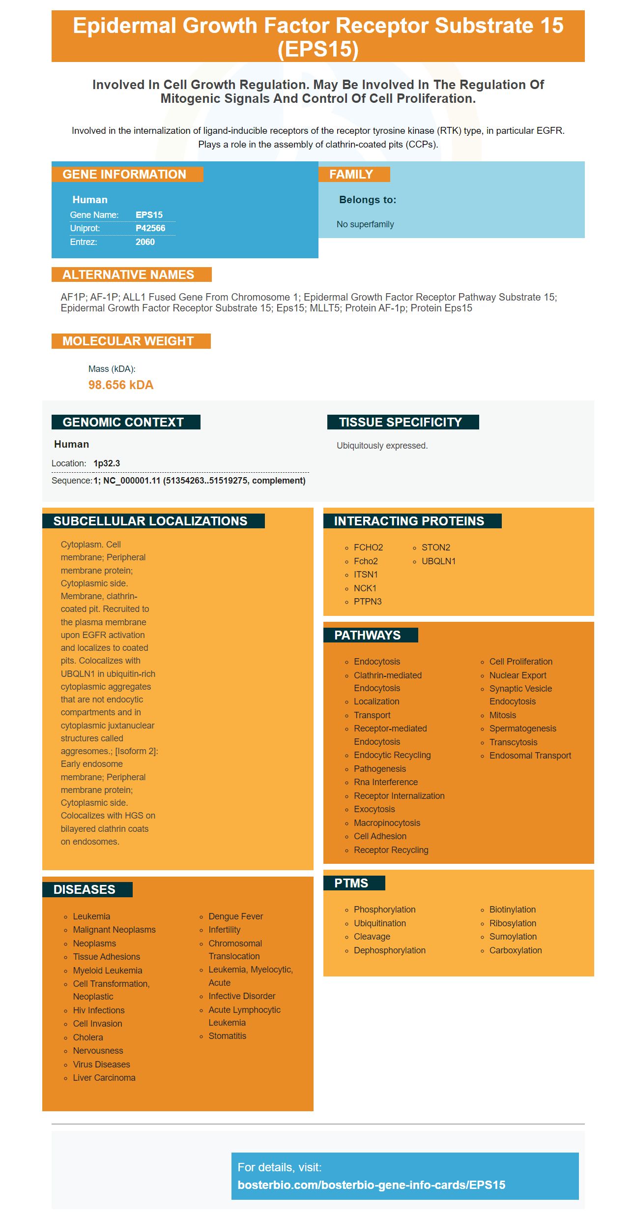
Facts about Epidermal growth factor receptor substrate 15.
Involved in the internalization of ligand-inducible receptors of the receptor tyrosine kinase (RTK) type, in particular EGFR. Plays a role in the assembly of clathrin-coated pits (CCPs).
Human | |
---|---|
Gene Name: | EPS15 |
Uniprot: | P42566 |
Entrez: | 2060 |
Belongs to: |
---|
No superfamily |
AF1P; AF-1P; ALL1 fused gene from chromosome 1; epidermal growth factor receptor pathway substrate 15; epidermal growth factor receptor substrate 15; Eps15; MLLT5; Protein AF-1p; Protein Eps15
Mass (kDA):
98.656 kDA
Human | |
---|---|
Location: | 1p32.3 |
Sequence: | 1; NC_000001.11 (51354263..51519275, complement) |
Ubiquitously expressed.
Cytoplasm. Cell membrane; Peripheral membrane protein; Cytoplasmic side. Membrane, clathrin-coated pit. Recruited to the plasma membrane upon EGFR activation and localizes to coated pits. Colocalizes with UBQLN1 in ubiquitin-rich cytoplasmic aggregates that are not endocytic compartments and in cytoplasmic juxtanuclear structures called aggresomes.; [Isoform 2]: Early endosome membrane; Peripheral membrane protein; Cytoplasmic side. Colocalizes with HGS on bilayered clathrin coats on endosomes.
HIPA (high-affinity primary antibodies) are among the most powerful tools for your research. But the question is how to choose the most effective one? Here are some helpful tips and guides to optimize the experiment. If you're not sure what you should do with your experiment, go through the Boster Bio optimization guides to get the most value from your research. These guides will help you through each step of your research, from choosing an appropriate flow procedure to maximizing the results.
Recent studies have revealed several proteins that bind to EPS15 which is a protein found within nerve cells. These antibodies bind EPS15 principally in neurons, but they also bind to other EH domain-containing proteins. Researchers performed fluorescent transferrin-uptake experiments in triple-null HeLa 40 cells to measure EPS15 intracellular sequestration. They then performed an AP-2 assay based on plasmids to determine the amount of AP-2-positive co-coated clathrin structures.
Unlike other markers, EPS15L1 can be detected in all tissues, with the exception of fibroblasts. But, it is crucial for living in mice and plays an essential role for the nervous system. This is why high-affinity primary antibodies using the EPS15 marker will effectively detect Eps15 in the target cell. Researchers will be able develop high-affinity primary antibodies using the EPS15 marker that will enable them to recognize EPS15L1 in different tissues.
Since EPS15 is required to develop and maintain the health of red blood cells and their presence in cells is crucial to maintaining iron homeostasis. In mice that have conditional Eps15/Eps15L1 knockout (cDKO) mutations the transferrin receptor is damaged, leading to anemia that is characterized by microcytic hypochromic anemia.
The high-affinity primary antibodies that utilize the EPS-15 marker have been proven to be efficient in a variety of applications like immunoblotting ELISA and flow cytometry. They can also be used to identify proteins involved in disease and to analyze post-transcriptional modifications. These antibodies have been validated in a variety of research projects and have been produced by the collection of samples from a variety of species.
Another peptide that plays a very functional and versatile role during neural development is EPS15L1. Its function is not fully understood but studies of mammalian cell lines indicate that both proteins regulate endocytosis interchangeably. However despite the similarity of their roles, EPS15L1 has its own distinct function in the nervous system. Both proteins play an important part in the development of embryos.
Secondary antibodies are normally produced against the species of interest's immunoglobulins. If a goat is provided with mice IgG purified the animal will develop antibodies that recognize all IgG classes and fragments. However, if the goat is immunized using only mouse IgG1 antibodies then the goat will create only antibodies that are specific to this IgG subclass.
A well-established method is used to estimate the KD values of these antibodies. They are calculated using a method known as equilibrium dissociation (EDC), which determines the reversibility of bimolecular interactions. It also identifies an EPS15 marker that can be employed in primary antibodies. The EPS15 marker has been chosen because of its affinity towards human IgG. However the results weren't as clear as they could have been, because some antibodies were not useful.
The EPS15 protein is tripartite structure that measures 138-140 kDa in size. It is comprised of three evolutionary conserved EH protein–protein interaction domains as well as two Ubiquitin binding motifs. The amino terminal portion is mainly responsible for interacting with the EGFR, while the carboxy terminal region contains several copies of aspartate-prolinephenylalanine.
Steven Boster's story is fascinating. The man has been part of science for decades. Although he was referred to as an "lavatory scientist," he never considered himself an inventor. His inventions are used widely in the present, from ELISA kits to immunofluorescence imaging. Steven Boster's bio contains full details of his accomplishments. Here, we examine some of his most notable achievements.
PMID: 8183552 by Wong W.T., et al. The human eps15 gene, encoding a tyrosine kinase substrate, is conserved in evolution and maps to 1p31-p32.
PMID: 8134107 by Bernard O.A., et al. A novel gene, AF-1p, fused to HRX in t(1;11)(p32;q23), is not related to AF-4, AF-9 nor ENL.