This website uses cookies to ensure you get the best experience on our website.
- Table of Contents
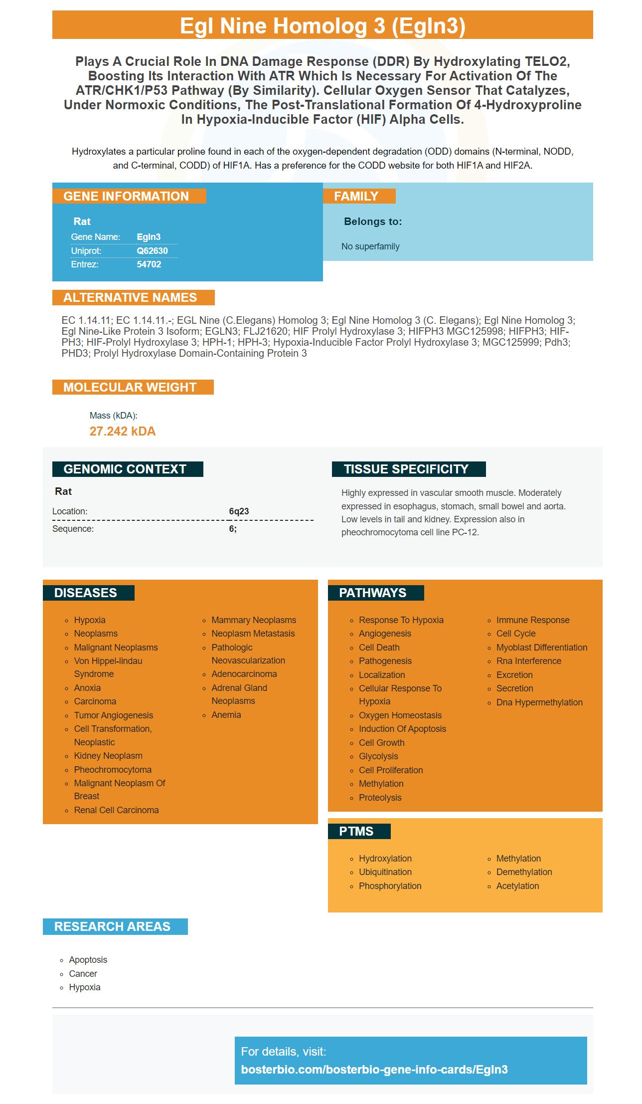
Facts about Egl nine homolog 3.
Hydroxylates a particular proline found in each of the oxygen-dependent degradation (ODD) domains (N-terminal, NODD, and C-terminal, CODD) of HIF1A. Has a preference for the CODD website for both HIF1A and HIF2A.
Rat | |
---|---|
Gene Name: | Egln3 |
Uniprot: | Q62630 |
Entrez: | 54702 |
Belongs to: |
---|
No superfamily |
EC 1.14.11; EC 1.14.11.-; EGL nine (C.elegans) homolog 3; egl nine homolog 3 (C. elegans); egl nine homolog 3; egl nine-like protein 3 isoform; EGLN3; FLJ21620; HIF prolyl hydroxylase 3; HIFPH3 MGC125998; HIFPH3; HIF-PH3; HIF-prolyl hydroxylase 3; HPH-1; HPH-3; Hypoxia-inducible factor prolyl hydroxylase 3; MGC125999; pdh3; PHD3; Prolyl hydroxylase domain-containing protein 3
Mass (kDA):
27.242 kDA
Rat | |
---|---|
Location: | 6q23 |
Sequence: | 6; |
Highly expressed in vascular smooth muscle. Moderately expressed in esophagus, stomach, small bowel and aorta. Low levels in tail and kidney. Expression also in pheochromocytoma cell line PC-12.
Many biological assays employ antibodies that react with HIF1A. This protein plays an important role in embryonic blood vessel formation, tumor angiogenesis, and pathophysiology for ischemic diseases. Boster Bio has developed these antibodies to detect HIF1A within animal samples. These antibodies can detect a wide range of species, including rabbit, mouse, and human.
HIF1A plays a vital role in cell response to hypoxia. It regulates gene transcription. It is highly expressed during the development of tumors, myocardial infarction, and wound healing. HIF-1a increases cell survival and proliferation genes. This extends cells' lives. Boster Bio's antibody is available in the Picoband catalog. It has been tested in both IHC as well as WB applications. It is compatible both with the mouse species and the rat species.
The HIF-1A marker cell lines were cultured with PX-478 which is a boster antibody that targets HIF-1A. Boster Bio has developed a method that targets the HIF-1A pathway at different cellular levels. This method is highly selective for this gene. It prevents the growth of cancer cells and decreases the number of angiogenic cell. Visit the BosterBio website for more information.
Boster bio HIF1A antibody, a highly select mAb, targets this protein on multiple levels. It inhibits HIF-1a Translation, Glucoxylase Activity and Deubiquitination. The antibody reduces the growth of cancerous cells and angiogenic ones. Boster Bio HIF1A antibody containing the EGLN3 mark has shown remarkable effectiveness.
HIF1A expression is frequently overexpressed in cancerous tumors and metastases. It is a key regulator of oxygen homoeostasis, and activated by tumor hypoxia. Hypoxia also stimulates VEGF production which supports tumor cell growth and invasion. Multiple studies have shown that tumors harboring EGLN proteins exhibit an array of characteristics, including increased angiogenesis. EGLN2 is closer to the TGFss pathway then EGLN3. Both proteins are positively correlated.
HIF1A plays an important role in the regulation of gene expression and cell response to hypoxia. It is highly expressed during tumor growth, myocardial infarction, and wound healing. HIF-1a is a protein that helps extend the life of cells by increasing expression of cell survival and proliferation genes. It also regulates gene expression across many organs including brain, liver and bone marrow. Boster Bio HIF1A antibody, based on EGLN3 marker, has excellent reproducibility. It can be used in many research fields.
Researchers were able to identify pancreatic cancer cells that had EGLN3 in their serum using the EGLN3 marker. This could allow doctors to choose the best treatment for their patients. It could also help patients determine if the disease has spread beyond their local area. The new antibody detects high levels HIF-1A. BosterBio HIF1A is fast-acting and highly specific, in addition to its high sensitivity.
The HIF1A protein, also known as hypoxia factor-1 alpha, is a key regulator of gene expression. It is expressed in high levels in many tissues such as embryonic and tumor angiogenesis, wound healing, or embryonic blood vessel formation. HIF1A stimulates cell survival and proliferation genes. This can extend cell life. Boster Bio has included the anti-HIF1A antibody in its Picoband catalog. The antibody is tested in various application including WB, IHC and other. The antibody reacts with both rabbit and mouse tissues.
Boster Bio's clinical team used BV-2 and PC-12 cells to evaluate the effects of Salidroside in a clinical study. The cells were cultured, treated with rhodiosin and tyrosol, then harvested using TRIzol reagent. The samples were reverse-transcribed into cDNA, and the cDNA was amplified using the ChamQ Universal SYBR-qPCR Master Mix and DNA-polymerase chain reaction.
EGLN3 hydroxylates HIF2A as well as PKM in hypoxia. These proteins are vital for glycolysis. By analyzing EGLN3, researchers can identify the underlying cause of hypoxia-induced diabetes and its therapeutic potential. With this knowledge, Boster Bio can further develop new drugs and vaccines that improve the health of their patients.
The HIF1A gene plays a key role in the response of cells to hypoxia and regulates gene expression. It is highly expressed during tumor growth, myocardial infarction, and wound healing. HIF-1a regulates cell survival and proliferation gene transcription, which extends cell life span. This antibody belongs to Boster Bio's Picoband catalog. It has been tested to detect IHC and WB reactions in rats and mice.
Researchers have discovered that HIF-1A can block glucoxylase-1 and inhibit tumor growth by targeting it. Boster antibodies have been validated in Western Blotting, Immunohistochemistry, and ELISA. As a result, they are a good choice for research. Boster bio also sells EGLN3 and other markers of RNA.
PMID: 8175725 by Wax S.D., et al. Identification of a novel growth factor-responsive gene in vascular smooth muscle cells.
PMID: 10386996 by Lipscomb E.A., et al. Expression of the SM-20 gene promotes death in nerve growth factor- dependent sympathetic neurons.