This website uses cookies to ensure you get the best experience on our website.
- Table of Contents
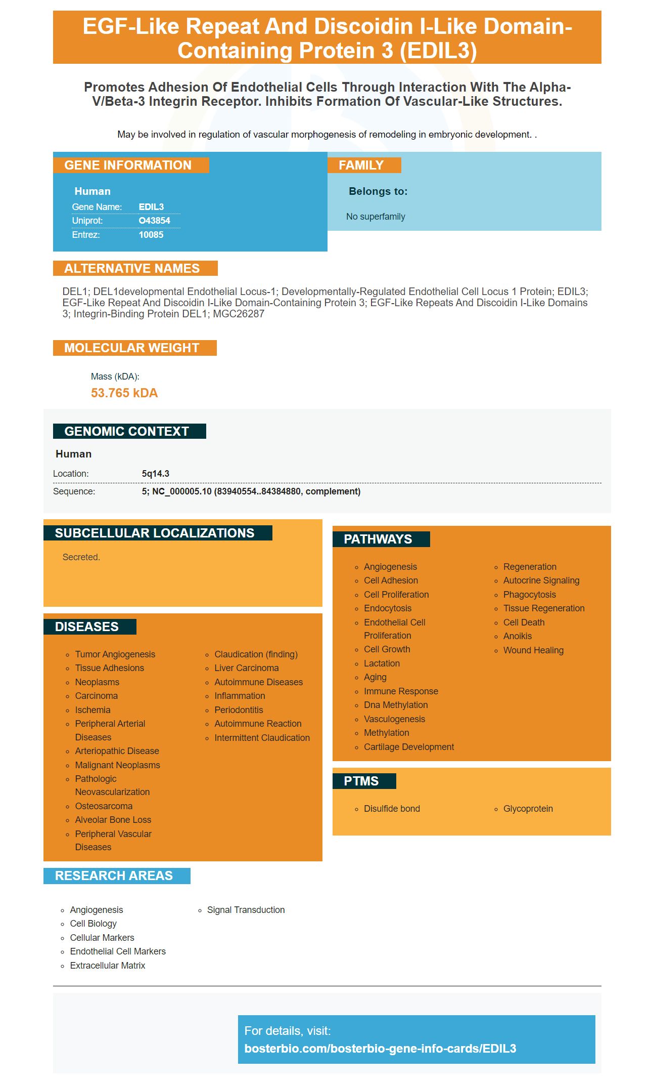

Facts about EGF-like repeat and discoidin I-like domain-containing protein 3.

May be involved in regulation of vascular morphogenesis of remodeling in embryonic development. .
| Human | |
|---|---|
| Gene Name: | EDIL3 |
| Uniprot: | O43854 |
| Entrez: | 10085 |

| Belongs to: |
|---|
| No superfamily |

DEL1; DEL1developmental endothelial locus-1; Developmentally-regulated endothelial cell locus 1 protein; EDIL3; EGF-like repeat and discoidin I-like domain-containing protein 3; EGF-like repeats and discoidin I-like domains 3; Integrin-binding protein DEL1; MGC26287
Mass (kDA):
53.765 kDA

| Human | |
|---|---|
| Location: | 5q14.3 |
| Sequence: | 5; NC_000005.10 (83940554..84384880, complement) |
Secreted.


Boster Bio Anti-EDIL3 Antibody reacts with Human EDIL3 Protein, catalog number A06273. This antibody can last up to a year if stored at -20degC. However, the antibody can also be stored at 4degC until it is used. It is important to avoid repeated freeze-thail cycles. You can also validate its quality against positive or negative samples.
EDIL3 - an extracellular matrix protein, plays an important role for angiogenesis and tumor progression. A poor prognosis is associated with a higher level of EDIL3. This study aimed to investigate the role EDIL3 plays in the progression to endometrial and other types of cancer. We also looked into the correlation between EDIL3 gene expression and microvessel count in lung squamous-cell carcinoma patients.
These results also show that EDIL3 levels in NSCCL cells are higher in SCLC cell lines with stem-like and mesenchymal characteristics. It is not clear how these cells influence anti-tumor immunity. CSCs are becoming increasingly important in NSCLC therapy. But, it remains a question: How can EDIL3 used to improve the prognosis in advanced NSCLC patients?
NSCLC cells had significantly higher EDIL3 levels than normal tissues. Moreover, the circulating EDIL3 protein was also found to be significantly upregulated in tumor cells. The study also revealed that EDIL3 gene expression in NSCLC patients correlated with survival time. This suggests that EDIL3 can be a therapeutic target to NSCLC.
Although the results of this study indicate that increased miRNA-145 expression levels are predictive of survival and response to erlotinib treatment, there are several limitations of this study. One limitation is the fact that the authors used only stage IIB patients and IIIA patients. This could not have been applicable to other centers or stages. These findings support the development of novel therapies to improve survival rates and treatment outcomes.
This study was conducted using two types of SCLC-21H cells and NCIH82 cells. All three cell lines were maintained in complete H-glucose DMEM with 10% FBS. After 48 h of culture, the conditioned medium was collected for analysis. Afterwards, SCLC cells were isolated and the PBMC cells were cultured in co-culture at a 1:1 ratio.
Three cell lines were used to test EDIL3 expression in NSCPC. SCLC-21H and NCI-H82 cells were both established in vitro. In the same study, we examined a relationship between adherence and stemness. In these cell lines, we observed that adherent cells gave rise to a subpopulation of loosely adherent cells, which were viable and remained in suspension.
Recent research found that EDIL3 levels in squamous cells carcinoma were higher than those in normal tissue. This study suggests that EDIL3 expression may be a source of this antitumor agent. In a study of murine Lewis lung cancer cells, EDIL3 levels were also higher. EDIL3 amplification in murine Lewis lungs carcinoma cells led to an increase in vimentin and a decrease in E-cadherin.
Low EDIL3 levels in squamous-cell carcinoma are associated to poor clinical outcomes. This is also linked to the tumor size. The gene is associated to increased vascularization in various types of tumors including osteosarcoma, squamous cell carcinoma, and osteosarcoma. Gene therapy that induces apoptosis and inhibits tumor growth may improve patient outcomes. This study does not directly address EDIL3's role in squamous-cell carcinoma. However, it suggests that epigenetic modifications could affect cancer cells.
HCC is a complex process that involves EDIL3. It is highly expressed by patients with this disease. The protein is linked to tumor size and formation of thrombuses in the portal vein. EDIL3 promotes anchorage-independent growth by ligating integrins and inhibiting anoikis. It also stimulates integrin signal pathway in tumor cells.
EDIL3 ligand is important for aV -coupled integrins. Recent studies have shown EDIL3 can promote the vascularization and growth of cancer cells. This protein could therefore be a target for antiangiogenic therapy. Therefore, further research is needed to understand its role in this cancer type.
The authors performed an IHC test using three different HCC line cell lines. Using a phalloidin FITC (phalloidin-FITC) solution in the culture medium, EDIL3 could be stained. After this procedure, the cells were seeded onto a plate coated with 0.5% methyl cellulose. To measure EDIL3 expression in HCC, cells were grown in serum-free medium for 48 hours. A second experiment used the same technique but used serum-free media.
This study revealed that EDIL3 expression levels were significantly higher in tumor tissues than in nontumor tissues. Furthermore, CDCA7 copy number amplification is associated with the presence of tumor cells. Correlation analysis using GSE53625 Data confirmed this relationship. This study also confirms the positive correlation between expressions and copy numbers amplification.
Previous studies have shown an association between EDIL3 gene expression and mesenchymal phenotypes, but no direct correlation. Although tumor angiogenesis can be associated with high EDIL3 gene expression, no studies have assessed the role of EDIL3 on NSCLC prognosis. The current study suggests that EDIL3 might play a role in enhancing this cancer's angiogenesis.
EDIL3, or DEL-1, is a secreted extracellular matrix protein that regulates cellular growth, differentiation, adhesion, and apoptosis. It also inhibits immune-cell recruitment to an inflammation site. It is important for the progression of different types of adenocarcinoma, because inflammation contributes invasive characteristics to transformed cells. A tumor's microenvironment can promote metastasis from other organs. The expression levels of EDIL3 vary depending on the type of cell and the stage at which the tumorgenesis occurred.
Interestingly, EDIL3 gene expression levels in adolescent or adult patients were significantly associated to survival. Patients with adenocarcinoma had a higher overall survival rate than those with higher levels of EDIL3. However, EDIL3 expression wasn't associated with adenocarcinoma characterized with high levels microvessels. This could be due epigenetic modifications. In this instance, it is still necessary to identify the exact role that EDIL3 plays in adenocarcinoma.
Interestingly, EDIL3 gene expression levels were significantly higher for HCC than for NL and CL. It was also discovered that EDIL3 levels are associated with the prognosis in patients with HCC. Quantitative real-time PCR (qRTPCR) was used to measure the protein level. Moreover, the relative mRNA levels of EDIL3 were higher in HCC than in NL and CL.
In the current study, 83 patients suffering from gastric cancer were included. PCHNT specimens taken from the tumor margin were removed and examined for normal or dysplastic cells. The specimens were frozen in liquid nitrogen and kept at -80C until western blot analysis. The clinicopathological characteristics for gastric cancer patients were correlated to EDIL3 expression levels.
The authors confirmed the regulation of EDIL3 in a qPCR assay that identified EDIL3-binding MIR-137. Furthermore, they found that knockdown of EDIL3 inhibited cell invasion, migration, and EMT. They also concluded EDIL3 knockdown affected cell proliferation and migration. These results suggest that EDIL3 is essential for tumor development.
PMID: 9420328 by Hidai C., et al. Cloning and characterization of developmental endothelial locus-1: an embryonic endothelial cell protein that binds the alphavbeta3 integrin receptor.
PMID: 22601780 by Schurpf T., et al. The RGD finger of Del-1 is a unique structural feature critical for integrin binding.

