This website uses cookies to ensure you get the best experience on our website.
- Table of Contents
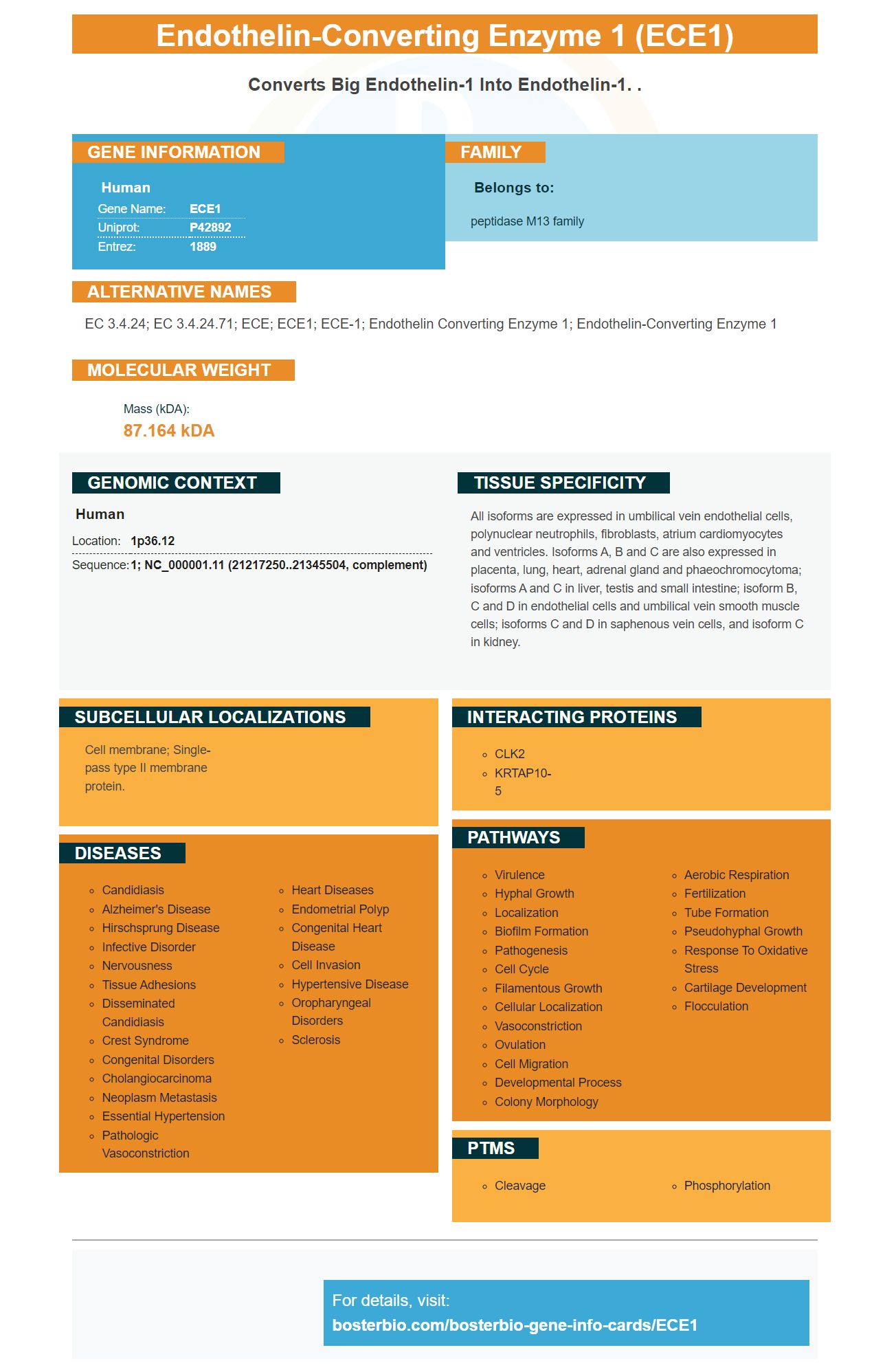
Facts about Endothelin-converting enzyme 1.
Human | |
---|---|
Gene Name: | ECE1 |
Uniprot: | P42892 |
Entrez: | 1889 |
Belongs to: |
---|
peptidase M13 family |
EC 3.4.24; EC 3.4.24.71; ECE; ECE1; ECE-1; endothelin converting enzyme 1; endothelin-converting enzyme 1
Mass (kDA):
87.164 kDA
Human | |
---|---|
Location: | 1p36.12 |
Sequence: | 1; NC_000001.11 (21217250..21345504, complement) |
All isoforms are expressed in umbilical vein endothelial cells, polynuclear neutrophils, fibroblasts, atrium cardiomyocytes and ventricles. Isoforms A, B and C are also expressed in placenta, lung, heart, adrenal gland and phaeochromocytoma; isoforms A and C in liver, testis and small intestine; isoform B, C and D in endothelial cells and umbilical vein smooth muscle cells; isoforms C and D in saphenous vein cells, and isoform C in kidney.
Cell membrane; Single-pass type II membrane protein.
Steven Boster (a Chinese immunologist) developed his first product late in 1990. This earned him "he who converts science to the lavatory" He later went on to develop several different products, including IHC and hundreds of primary antibodies. Boster was China's biggest antibody catalog company during the late 90s. Later, he patented PicoKine (tm), his proprietary ELISA technology. PicoKine(tm), a proprietary platform that delivers high-sensitivity ELISA kits to many diseases, uses trade secrets.
ECE1 can be described as a type II integral cell membrane protein. It contains a short N terminal cytoplasmic trail, an extracellular hemophobic domain, as well as a zinc-binding motif. There are four distinct ECE1 human versions. All of them are similar, with the exception of their C-terminal amino acids sequences. The specific targeting domains of each ECE1 isoform and their intracellular compartments are what distinguish them.
Substance P is destroyed in many cell types by the ECE1 proteins, including neuronal and neural cells. The protein has four isoforms in mice and humans. The isoforms share an external catalytic domain and have distinct N -terminal cytoplasmic trail regions. The signal sequence in the ECE1 ECE1 recombinant is involved with secretion.
ECE1 is a member of the peptidase Family M13 and is involved in the proteolysis endothelin precursors. They are not intended for diagnostic procedures or resale. However, the protein has become a key marker for diagnosing neuronal injury. This enzyme is part the HUGO nomenclature.
The ECE1 marker is expressed by the hyperactive Ahr1 genetic. Ahr1 binds with the promoters for CFR genes. This promoter-binding activity leads to increased expression and toxicity of CFR genes. Ahr1 also triggers the transcription and expression of CFR genes. Ahr1 mutations can result in a decrease or absence of CFR genes.
ECE1's ability to convert big-endothelin-1 and endothelin-1 has biological significance. Defects in ECE1 result in Hirschsprung disease, a rare disorder that causes both cardiac and autonomic dysfunction. ECE1 is a member of the peptidase family, M13. There are four different isoforms. Each one is distinguished by a distinct signal sequence and secretes an ectodomain.
Studies have shown that different types and levels of atherosclerosis have an increased level of the ECE-1 marker. In the early stages of atherosclerosis, the ECE-1 marker is also present. Inflammation is associated with upregulation of the ET-1 system, which could be a consequence of increased production of active ET-1. In addition, ET-1 may be responsible for regulation of vascular tone.
Double immunolabeling with the ECE-1 antibody intimal tissue confirms the presence and extent of ECs. In all but one case, ECE-1 immunoreactivity was present in a subset of ECs and intimal VSMCs. The ECE1-positive cells were associated with the nucleus. The result is a positive signal for ECs. There are many reasons for this marker's presence.
One of these is ECE-1 which promotes the internalization CGRP at the cell surface into endosomes. Consequently, it has a protective role in preventing fibrosis and inflammation. It has been implicated in pulmonary diseases and may be a marker to CGRP. It is also involved the regulation M2 macrophages and inflammatory disease.
ET-1 immunoreactivity in inflammatory and fibrotic areas was observed in 15 of fifteen cases. ECE-1 was also detected in the media underneath the plaques. Double immunolabeling found that ET-1 was expressed only in 15% luminal ECs. The remaining 15% were ECE-1+. Double immunolabeling confirmed ET-1 expression in macrophages. Despite this finding, ET-1 immunoreactivity could be detected in areas of plaque that had chronic inflammation. ET-1 immunoreactivity was also found in both extracellular (and intracellular) areas.
The ECE1 marker is involved in the proteolytic process of endothelin precursors. These precursors are then converted into biologically active proteins. ECE1 can be used to evaluate the severity and risk of endothelial disease, monitor drug-induced liver injury, and assess the risk of developing heart disease. Clinical applications of the ECE1 marker have several advantages.
The ECE1 protein can be expressed by many cells in the lung. It is mainly produced mostly by M2 macrophages. They are crucial for lung transformation. Its elevated levels have been linked with decreased inflammation and progression into fibrosis. ECE1 levels that are higher have been associated with better lung function. These effects suggest ECE1 is beneficial in the detection of lung disease.
Although the promising results are encouraging, more research is needed to establish clinical use of the ECE1 marker. The strain-to-strain ratio is an important factor in determining the ECE1 gene. ECE1 should be used in a strain-to–strain manner to identify the pathogens present at a particular location. This would ensure the correct treatment.
The ECE1 gene is abundant in different human arteries at different stages of atherosclerosis. In addition, the ECE-1/ET-1 system has been linked to chronic inflammation. ECE-1-positive cells were found in the inner lining, which is lower than the levels under fatty streaks or CPs. This suggests that ECE1 may be involved in controlling cell growth, vascular tone and inflammation.
The ET-1 marker is used to measure the invasiveness in ovarian cancer. In one study, it has been shown to decrease ET-1-dependent p44/42 mitogen-activated protein kinase activity and reduce the invasiveness of ovarian cancer cells. These encouraging results could lead to a novel anticancer treatment. The ECE1 gene is a promising candidate for clinical use, but it is important to understand its underlying mechanisms and clinical applications.
The ECE1 gene was also studied in mice. The mice challenged with the 3kbVO strain showed similar ECE1 immunoreactivity with the WT allele, while the TVO strain expressed 529L. Double immunolabeling confirmed that ECE1 expressing mice had less PMN recruitment, tissue destruction, and IL-1b release. The results suggested that ECE1 variation may have contributed to the decreased vaginal immunitypathology.
Molecular fusions could increase the diagnostic accuracy of one biomarker. Combining total PSA and free form PSA can improve cancer-specificity. CA125 and HE4 can be used as biomarkers for tumors. These two biomarkers are routinely used to detect late stage ovarian cancer and monitor treatment. Combination studies can improve the clinical utility and efficacy of this biomarker.
PMID: 7695628 by Yorimitsu K., et al. Cloning and sequencing of a human endothelin converting enzyme in renal adenocarcinoma (ACHN) cells producing endothelin-2.
PMID: 7864876 by Shimada K., et al. Cloning and functional expression of human endothelin-converting enzyme cDNA.
*More publications can be found for each product on its corresponding product page