This website uses cookies to ensure you get the best experience on our website.
- Table of Contents
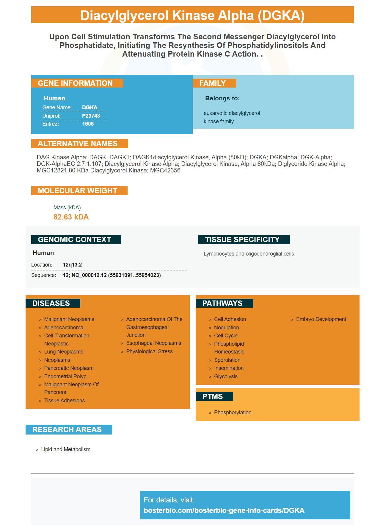
Facts about Diacylglycerol kinase alpha.
Human | |
---|---|
Gene Name: | DGKA |
Uniprot: | P23743 |
Entrez: | 1606 |
Belongs to: |
---|
eukaryotic diacylglycerol kinase family |
DAG kinase alpha; DAGK; DAGK1; DAGK1diacylglycerol kinase, alpha (80kD); DGKA; DGKalpha; DGK-alpha; DGK-alphaEC 2.7.1.107; diacylglycerol kinase alpha; diacylglycerol kinase, alpha 80kDa; Diglyceride kinase alpha; MGC12821,80 kDa diacylglycerol kinase; MGC42356
Mass (kDA):
82.63 kDA
Human | |
---|---|
Location: | 12q13.2 |
Sequence: | 12; NC_000012.12 (55931091..55954023) |
Lymphocytes and oligodendroglial cells.
If you are searching for the DGKA markers, you have come to the right spot. This article will discuss the advantages and uses of the DGKA Cell-Based ELISA Kit. Below are some other uses for the DGKA marker. These include: * Monitoring and screening for the effects on DGKA expression cells by activators and treatments.
The DGKA improver is a target genetic gene for a novel kind of therapeutic drug. The DGKA enhancer can be genetically engineered to produce animal models with altered DGKA levels. This would make it possible to bridge the gap between bench and bedside drug discovery. You can modify the DGKA enhancer to block the activity of many important pathways in your body. It has been demonstrated that DGKA Enhancers can reduce the activity a specific enzyme.
The DGKA promoter is located in the genomic region. DNA methylation levels differed between the three NHDF cell line lines. L1-L4 fibroblasts had low levels while h2-H2 cells had high levels. h2 and h2 cells also had higher levels the methylation mark, CpG_6, located at the first EGR1 bind site.
Interestingly, increased DGKA gene expression was associated with DNA demethylation of DGKA enhancer. Demethylation also of the downstream control regions and upstream promoter resulted in a higher DGKA level. Treatment with cumate resulted in enrichment of H3K27me3, suggesting demethylation of DGKA enhancer promotes transcript.
Using a multidimensional scale approach, JQ1 & CBP30 prevented DGKA induction upon irradiation. These inhibitors had a limited effect on p300. DGKA enhancers cannot be used to cure cancer. It is important that we identify the mechanism that suppresses cancer. There are many ways to stop cancer from occurring.
Radiation-induced fibrosis can be caused by alterations of DGKA gene expression. Radiation-induced profibrotic marker mutations are associated with alterations at the methylation level of the DGKA promoter. Small molecules that target DGKA Enhancer can reduce fibrosis risk. These small molecules may play an important role for cancer prevention.
Other regions of fibroblast genome may also be implicated in the DGKA boostr. The genes COMP, PDGFA and ADRA2A are all involved in cancer. Most of these pathways involve DNA replication and/or repair. Nine pathways were shared between the sgC & sgE1 cells. Multi-factorial analysis by DGKA Enhancer of EGR1-binding revealed at least 272 DEGs at DGKA Enhancer in sgE1 Cells. 144 of these genes are upregulated and one hundred and eighty four percent are downregulated.
The DGKA enhancer inhibits irradiation-induced expression of COL1A1 and DGKA. GSK126, EPZ6438 inhibit radiation-induced DGKA production. The DGKA enhancer reduces this expression by increasing activity of COL1A1 in the liver and GSK126. It also suppresses COL1A1.
The DGKA ELISA Kit Cell-Based is highly sensitive and easy to use for screening a wide range targets, including DGKA. Scientists can monitor changes in phosphorylation status for a protein in cell-based assays with this kit. Scientists can also use the kit to determine the effects on DGKA levels of different activators or inhibitors. This kit is available to researchers of all disciplines around the world.
The DGKA cell-based ELISA kit is designed to detect levels of the cancer biomarker DGDKA in human cell culture, tissue, and cultured cells. The ELISA test kit has been validated to detect a wide array of samples. Validation images and procedures can be provided upon request. Picoband secondary antibody is polymer based and takes about 30 minutes for IHC. This antibody is backed up by knowledge about immunogen design and technical support from Sanbio distributor BeNeLux.
The development of a highly sensitive and specific method to detect high-affinity prima antibodies using the DGKA marker was long anticipated. The DGKA marker, a novel molecule with a high affinity for the antigen CD20, allows the detection of antibody pools that are specific for COVID-19. These antibodies recognize both CD20 and CD8 antigens. Using this method, we can identify high-affinity antibodies from patients suffering from different types of disease.
Surface plasmon imaging is a method to measure the specificity of antibodies and their binding strength. To determine the specificity of the antibodies, we used a RBD library of peptides. After taking measurements, we used the peptide epitope mapping method to determine the specificity of antibodies. The results of these measurements are presented in the next sections. The boxplots below show the results. The median value can be represented as a boxplot. The boxplot contains the p25 (and p75) values. The SPRi U value was calculated using the Mann-Whitney U test. Multiple comparisons could be corrected using Bonferroni–Holm.
We developed a peptide database that included 24 peptides that have known binding motifs. The four-amino acid overlap motif was selected and the exposed moieties. We highlighted in green known binding motifs to help us identify them. The Ig dRU signals were then log10 transformed to reveal their dRU signal. Next, we ran a secondary antibody detection assay.
The results showed a significant difference in the kd response between patients with acute and critically ill patients. This difference could not be explained by gender, body mass, or age. The kd responses to peak LDH and CRP levels did not show statistical significance. It was interesting to note that the kd response was significantly greater in patients with severe and hospitalized disease. However, the IgG response remained significant when the dpo was removed for less than 10 days.
PMID: 2175712 by Schaap D., et al. Purification, cDNA-cloning and expression of human diacylglycerol kinase.
PMID: 8180475 by Hart T.C., et al. Assignment of the gene for diacylglycerol kinase (DAGK) to human chromosome 12.