This website uses cookies to ensure you get the best experience on our website.
- Table of Contents
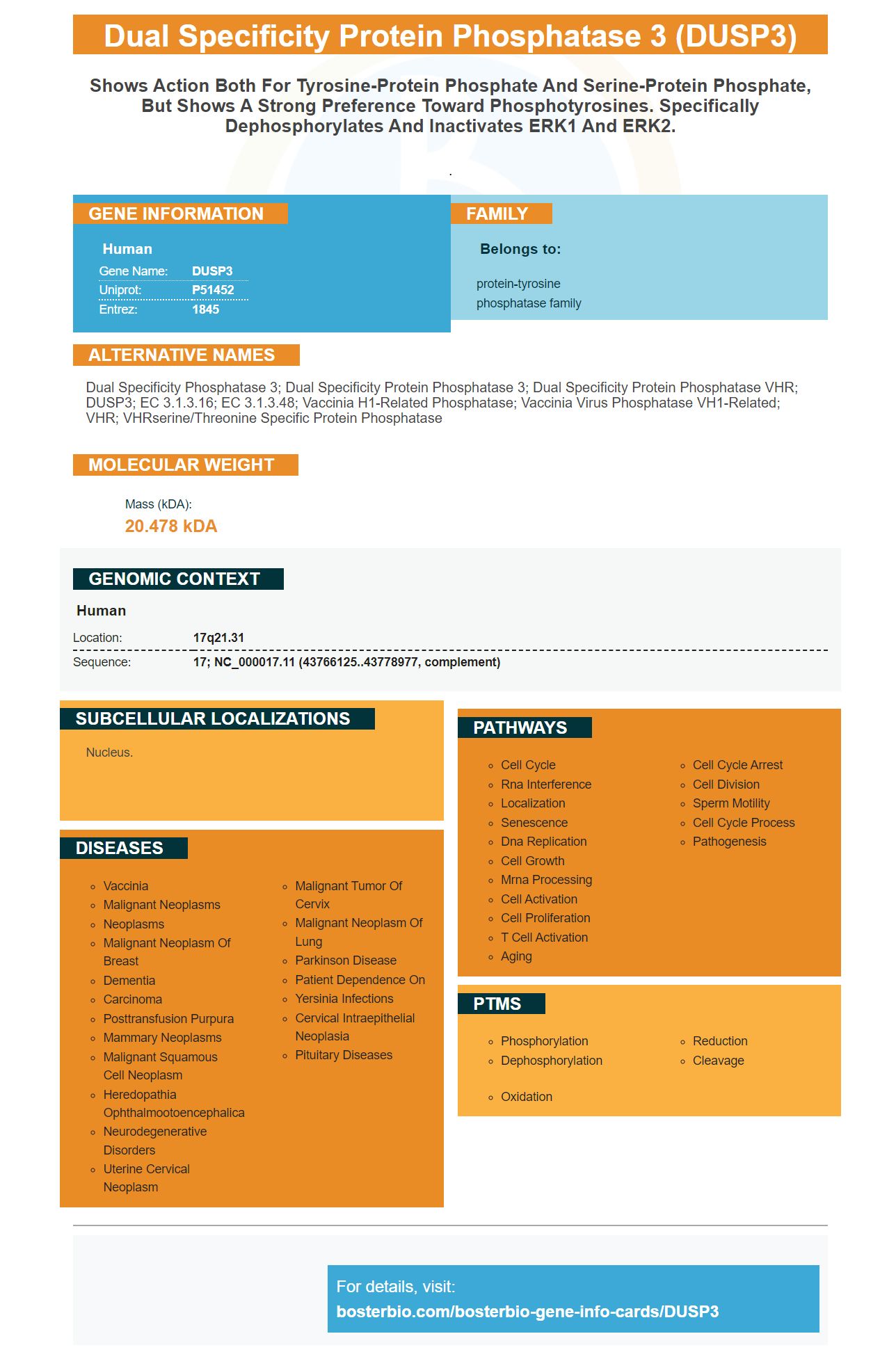
Facts about Dual specificity protein phosphatase 3.
.
Human | |
---|---|
Gene Name: | DUSP3 |
Uniprot: | P51452 |
Entrez: | 1845 |
Belongs to: |
---|
protein-tyrosine phosphatase family |
dual specificity phosphatase 3; dual specificity protein phosphatase 3; Dual specificity protein phosphatase VHR; DUSP3; EC 3.1.3.16; EC 3.1.3.48; Vaccinia H1-related phosphatase; vaccinia virus phosphatase VH1-related; VHR; VHRserine/threonine specific protein phosphatase
Mass (kDA):
20.478 kDA
Human | |
---|---|
Location: | 17q21.31 |
Sequence: | 17; NC_000017.11 (43766125..43778977, complement) |
Nucleus.
The DUSP3 indicator is one of the most popular markers for monitoring proliferation. Boster Bio offers high-affinity primaries against DUSP3 to aid researchers in a variety opportunites. Boster antibodies have been highly cited in the scientific literature for 25 years and are well-validated on immunohistochemistry, Western blotting, and ELISA. This is why Boster products have been trusted by researchers.
Boster Bio, a world-renowned company, offers high-affinity primary antibodies to DUTP3. Their reagents are highly precise and validated for specificity as well as high affinity. They also reward the product credit reviewers who are the first to review the products. They also reward scientists all over the world for their contributions to scientific communities. DUTPase a key protein involved in tumorigenesis.
DUSP3 is a member of the dual specificity protein phosphatase familly. The protein is composed of 185 amino acid and does not contain a docking site or targeting domain. It is encoded by the gene DUSP3/Dusp3. Although DUSP3 has been implicated with tumorigenesis, its function remains largely unknown. This antibody targets DUSP3 with high affinity. This is essential for accurate quantification.
DUSP3 Downregulation does not affect cJun phosphorylation, or JNK activitiy. It did not affect the phosphorylation and activity of ERK1/2. Boster Bio supplies high-affinity primary antibodies against DUSP3 because DUSP3 is an important part of the cell's immune system.
DUSP3 is a component of angiogenesis. In addition, this protein contributes to growth factor-induced sprouting. By blocking DUSP3 expression in angiogenesis-inducing cells, it blocks the growth of the angiogenesis process. This is a significant step towards the prevention of cancer.
DUSP3 immunoreactivity has been tested using a variety of commercially available reagents. Boster Bio's anti DUSP3 Picoband (tm) antibody (catalog#A06135) reacts with DUSP3 cells from Human and Mouse. The product contains both the recombinant Human DUP3 protein and Trehalose.
The researchers used mice to study the role of DUSP3 in tumor development. In a mouse model, mice without DUSP3 showed no signs or vascular deformation. This suggests that its function could be redundant. Because several DUSPs are substrate-specific, it is difficult to assign a definite physiological function. Conditional knockout mice lacking DUSP3 can't develop neovascularized tumors.
These experiments were performed on mice that did not have DUSP3 and were injected with 0.5 mLs of Matrigel containing 250 ng/mL bFGF. The solution was diluted with heparin. The plugs were then dissected and the hemoglobin content was measured using Drabkin’s reagent. The results were then measured using Imaris software.
The DNA damage reaction refers to cell repair and DNA phosphorylation (H2AX at Se139). This response has also been demonstrated in different cell systems. DUSP3 dephosphorylation decreases the stability of NUCL/NPM and leads to increased phosphorylation ARF and HDM2. DUSP3 is also crucial in the regulation and transcriptional activity for the p53 protein.
DUSP3 is overexpressed in cervical cancer cells, localized in the nucleus. Its knockdown results is a decrease of CDC2 or CDK4 and induces cellular senescence. DUSP3 is essential for the inhibition of androgen-induced apoptosis. This action is initiated by DUSP3 which activates MAPK.
In recent studies, researchers found that DUSP3 is involved in radioresistance and sensitivity in human tumoral cells. Radiotherapy has long been concerned about the proliferative responsiveness to radiotherapy-related cell lines. Two human cell lines, MeWo (and HeLa) were studied to see if they were more sensitive than others to gamma radiation.
These studies proved that gamma-radiation and DUSP3 impeded one another's DNA repair. After being exposed to 5 Gy of Gamma radiation, the cells were examined using flow cytometry. IR caused DNA double strand break, but cellular senescence resulted from the loss of DUSP3. Both mechanisms could be suppressed with a single treatment.
These findings indicate that DUSP proteins contain distinct catalytic domains which bind to Tyr(P),-peptides. These enzymes have substrates that vary in length from 171 amino acids in Vh2 and 380 in Cdc25A. These amino acids have the highest kcat/Km compared to all DUSP protein sequences. Further research is needed to determine the exact mechanism of cell growth in these cells.
DUSP3 inhibits ATM, ERK1/2 and JNK. DUSP1 has been the first DUSP found in mammals. DUSP3 is responsible for dephosphorylating ERK1/2, p38 and JNK. DUSP22 is also required to activate the JNK signaling pathway fully and is required for DNA damage when ATM or DUSP3 inhibitor is present.
DUSPs are small protein phosphatases with a relatively shallow active site. They can bind to all four tyrosines, and are characterized by a high binding affinity to phospho-Y-decapeptides. It has been suggested that DUSP3 is necessary for phosphorylation because of its low KD.
DUSPs play a crucial role in cellular signaling. DUSP3 falls under the DUSP13B family which also includes DUSP26, DUSP27, and DUSP13B. DUSPs have been implicated as a cause of several human cancers. However, their tumor-suppressing properties are not yet fully understood. DUSPs are present in most human tissues and can be found in tumor samples.
DUSP3 regulates cell cycle checkpoints and DNA repair pathways. It has also been implicated with cell-cycle arrest. It interacts directly with the nucleophosmin Protein (NPM). The presence of specific antibodies has shown UV-stress induced tyrosine phosphorylation in DUSP3 DUSP3 knockdown results are early nucleolus exit by NPM and ARF proteins as well as disruption of the HDM2-p53 interplay in the nucleoplasm.
DUSP3 is able to specifically dephosphorylate three tyrosine atoms and can also decrease phosphorylation levels when Na3VO4 is present. This study demonstrates that the DUSP3 protein gene can be expressed in a biological material. Three independent experiments are presented, with each showing four immunoblot results. The data in this paper are not conclusive, but they are representative of the literature.
UV radiation phosphorylates p53 Ser15. Similarly, dephosphorylation DUSP3 results in increased p53 activity. In control cells, the DUSP3 gene is silenced, whereas the expression of the DUSP3 marker increases in the DUSP3-silenced group. Therefore, DUSP3 plays a critical role in the development of human cancers.
Several types of tumor cells have the DUSP3 genes. The marker has a high affinity in biological samples for DUSP3. However, its high affinity for this enzyme is not reflected in its native form. DUSP3 expression is found in the cellular nuclear plasm of a tumor-cell. Interestingly, tumor cells expressing DUSP3 are more active than normal human cells.
The DUSP3 marker can be used to evaluate cell proliferation. This marker is expressed on the cell surface. One study examined DUSP3 expression in BL-21 cells. The cells were stained using centrin-3 antibodies. Half of the cells had two foci while the other half had more than two foci. Dusp3 knockdown cells were named shDUSP3 and were derived from the DUSP3 gene from Life Technologies.
A total lysate from MRC-5 shDUSP3 cells was prepared after UVC treatment to determine the DUSP3 level. Phosphorylase Y decapeptides have a high binding affinity for DUSP3-3WT. The DUSP3–WT protein has a lower KD than the full-length NAPM, but it is necessary to phosphorylate.
Proliferation refers to cell division and synthesis. Proliferation is defined by the presence of proteins in proliferating cells, but not in non-proliferating ones. We need primary antibodies to detect these proteins in order to study cell proliferation. These antibodies can be used in flow-cytometry and can be used to analyze cell proliferation. If you have a particular protein or antigen, you may be able to create a flowcytometry panel using this marker.
DUSP3 dephosphorylates the NPM, which drives the checkpoints during the cell cycle and the DNA repair pathways. The NPM translocation into the nucleoplasm occurs within three hours of DUSP3 knockout cells expressing the DUSP3 protein. Furthermore, the DUSP3 knockdown caused nucleoplasmic NPM to be detected in NS cells six hours after UV treatment.
DUSP3 knockdown cellular cells exhibit increased phosphorylation p53(Ser15), as a result of UVC radiation. HDM2 is also recruited to the nucleoplasm, where it is sequestered towards the nucleoli. In shDUSP3 cells, p53(Ser15) phosphorylation peaks at 3 h and three hours after UVC exposure, while it peaks at six hours in NS cells. Similarly, DUSP3 knockdown cellular cells show spatiotemporal phosphorylation of p53 and ARF. Although these markers are known to interact, we still don't know how.
The DUSP3 gene can be expressed in a variety of cell types and plays important roles during cell cycle phases. Dusp3 not only regulates cell proliferation but also plays a key role in mitosis, spindle assembly, and other functions. Its knockdown expression is useful for monitoring various types of tumors, including cancers. It can also be used in cell therapy to target cancerous tumors.
PMID: 1281549 by Ishibashi T., et al. Expression cloning of a human dual-specificity phosphatase.
PMID: 10224087 by Todd J.L., et al. Extracellular regulated kinases (ERK) 1 and ERK2 are authentic substrates for the dual-specificity protein-tyrosine phosphatase VHR. A novel role in down-regulating the ERK pathway.