This website uses cookies to ensure you get the best experience on our website.
- Table of Contents
4 Citations 6 Q&As
1 Citations 9 Q&As
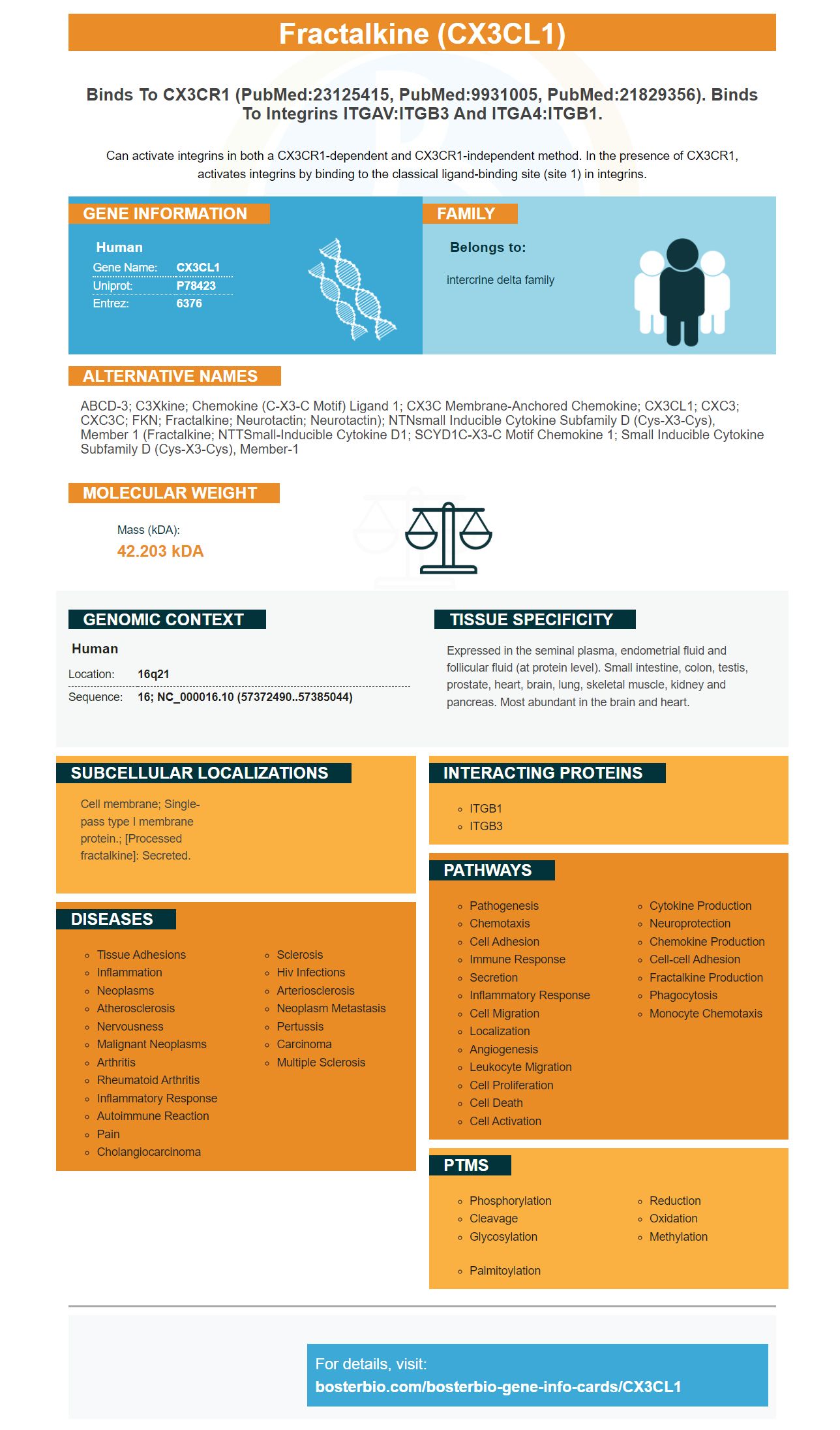
Facts about Fractalkine.
Can activate integrins in both a CX3CR1-dependent and CX3CR1-independent method. In the presence of CX3CR1, activates integrins by binding to the classical ligand-binding site (site 1) in integrins.
Human | |
---|---|
Gene Name: | CX3CL1 |
Uniprot: | P78423 |
Entrez: | 6376 |
Belongs to: |
---|
intercrine delta family |
ABCD-3; C3Xkine; chemokine (C-X3-C motif) ligand 1; CX3C membrane-anchored chemokine; CX3CL1; CXC3; CXC3C; FKN; Fractalkine; Neurotactin; neurotactin); NTNsmall inducible cytokine subfamily D (Cys-X3-Cys), member 1 (fractalkine; NTTSmall-inducible cytokine D1; SCYD1C-X3-C motif chemokine 1; small inducible cytokine subfamily D (Cys-X3-Cys), member-1
Mass (kDA):
42.203 kDA
Human | |
---|---|
Location: | 16q21 |
Sequence: | 16; NC_000016.10 (57372490..57385044) |
Expressed in the seminal plasma, endometrial fluid and follicular fluid (at protein level). Small intestine, colon, testis, prostate, heart, brain, lung, skeletal muscle, kidney and pancreas. Most abundant in the brain and heart.
Cell membrane; Single-pass type I membrane protein.; [Processed fractalkine]: Secreted.
This article will explore the many applications of CX3CL1, an chemical chemokine ligand that has a CX3C motif. Its main functions are to promote cell adhesion to VBMECs and increasing NSCLC cell migration. Read on to learn more about its possible applications. You can then utilize it for your research.
This study shows that CX3CL1 facilitated cell migration and invasion through GEFh2/Src pathways. We further show that CX3CL1 increased the permeability of VBMECs by improving cell adhesion. We also report that CX3CL1 activates the ADAM17 chemokine receptor that is expressed by endothelial cells.
This chemokine-ligand binds VBMECs and is more abundant in vertebral bones, limb bones kidney tissues, and lung cells. It also facilitates the entry of NSCLC cell lines and increases their adhesion towards VBMECs. It is also a key player in the MAPK14/ADMA17 and NFKB pathways that allow for increased NSCLC cell invasion. Further the spinal NSCLC metastases were linked to increased expression of CX3CL1.
The red bone marrow of vertebrae has an unique blood sinus structure that is home to many cytokines, enzymes and hormones. Chemokines are small structurally related cytokines play critical roles in immune system and inflammation. CX3CL1 has been identified as an important mediator of tumor metastasis in research on cancer. Its increased expression in BMECs may be related to the greater tumor metastasis of HCC cells.
Additionally, Boster Bio CX3CL1 is an excellent anti-tumor effect product. It has been shown to decrease the movement of HCC cells when in co-culture with cells. Inhibition of ADAM17 significantly reduced this anti-tumor activity, which suggests that CX3CL1 enhances adhesion of cells to VBMECs.
As an soluble chemokine ligand CX3CL1 draws monocytes and T cells. Cell-bound CX3CL1 enhances the adhesion of leukocytes to activated endothelial cells. In addition, CX3CL1 interacts with CX3CR1 to activate its adhesive properties. This chemokine is found on the human 16th chromosome.
In the study the levels of CX3CL1 were determined by using a co-culture of A549 cells and VBMECs treated with conditioned media. The results of this study were compared to the results of the control group. The anti-inflammatory effects of the drug on MMP activity were also examined using co-cultured LFA-1 and the a549 cell line.
In the study, the mRNA and protein levels of CX3CL1 were increased in tumor bone compared to normal vertebral bone. These results support the hypothesis of CX3CL1 being a major player in spinal metastases. Alongside regulating the growth of tumors, CX3CL1 is also a booster of the immune system and fights inflammation.
CX3CL1 expression in lung cancer cells has been linked to the development of spinal metastasis. It is controlled by the cell adhesion protein ICAM-1. It is also linked to VBMECs, also known as vascular endothelial cells (VBMECs) and can enhance the invasion of tumor cells and adhesion. This feedforward loop helps in NSCLC spinal metastasis.
Human Fractalkine, a human protein has been found to bind to an anti-body. This ELISA examines the amount target bound to the sample. The antibody pair that is matched binds to the target-specific antibody and the detector antibody. The enzyme-antibody-target complex is then reacted with the sample in a sandwich-like format. The concentration of target in the original specimen determines the intensity of the signal.
VBMECs bone marrow-derived endothelial cell (BMECs), are an important component in the microenvironment of the marrow. They are the principal source for cytokines within cancellous bones. The CX3CL1/ICAM-1 expression in VBMECs was greater than that of LBMECs during the third passage. Western blotting analysis also indicated that VBMECs have more CX3CL1 expression than LBMECs.
We first confirmed the presence of CX3CL1 in cells to determine if it enhances cell adhesion to VBMECs. Cells were seeded in 24-well Transwell chambers and then treated with polybrene (6 ug/ul). The cells were then incubated with the PI3K and AKT inhibitors MK-2206-2HCI, for 6 days.
CX3CL1 and ICAM-1, an anticancer drug, increased the permeability to VBMECs. This was accomplished through the Src/GEFh2 pathway. CX3CL1 improved the permeability of VBMECs as well as facilitated NSCLC cell transendothelial migration. The researchers concluded that this drug may prevent NSCLC from metastasizing to new organs.
This study demonstrated that platelets play an important role in adhesion of NSCLC cells to VBMECs. The inhibition of CX3CL1 through genetic means reduced the amount of adherent cells within the control VBMEC groups. The increase in adhesion caused by platelets was halted through the genetic silencing CX3CL1. This suggests that platelets promote cell adhesion to NSCLC cells in a CX3CL1/ICAM-1-dependent manner.
CX3CL1 increases cell adhesion in VBMECs via interactions with CX3CR1 or ICAM-1/LFA-1. The interaction between CX3CL1 and ICAM-1/LFA-mediated cellular adhesion to VBMECs creates a virulent feedback loop that facilitates the metastasis of NSCLC.
CX3CL1 is a cadherin-like protein which connects adjacent cells via metal ion-dependent homophilic interactions. It is part of the extracellular matrix, also known as the ECM. Cell adhesion proteins play a crucial role in the organization of epithelial morphogenesis, the cytoskeleton, and cell structure. The hallmark of the progression of cancer is the loss of cadherin expression.
ICAM-1/LFA-1 protein expression is associated with an increase in metastasis in NSCLC spinal metastases. The number of cells expressing ICAM-1 was increased in VBMECs in co-cultures. NF-kB inhibition decreased this effect. Additionally, these findings support a link between CX3CL1 and tumor progression.
The production and release of CX3CL1 by NSCLC cells is controlled by MAPK14 and NF-kB signaling. ADAM17 was upregulated in VBMEC-co-cultured h2975 and A549 cells. Additionally, PI3K/AKT inhibition slowed the increase in CX3CL1 in VBMECs through reducing the amount of ADAM17.
A new study reveals that the increasing expression of the CX3CL1 marker assists in NSCLC cell migration and the extravasation into vertebral cancellous bone. The results suggest novel signaling pathways that could be targeted for the development of early prevention and treatment strategies for spinal metastasis caused by NSCLC. The research suggests that CX3CL1 expression is higher in the vertebral and lung bone.
The researchers will next examine how CX3CL1 expression influences lung cancer cell migration. They will then transplant 500 ng CX3CL1-expressing A549 cells into mice without nucleus. They will also analyze immunoNF-tochemistry results to find out the relationship between CX3CL1 expression and malignant grade. They will also determine whether CX3CL1 is linked to other proteins, like Bcl2 and PCNA.
This study further confirmed that a high level of expression of CX3CL1 caused lung cancer cell migration. CX3CL1 also triggered matrix-metalloproteinase MMP-3 and MMP-9 to be elevated in patients with intrahepatic-cholangiocarcinoma. These factors directly correlated with poor prognosis. The CX3CL1 protein plays a crucial role in cancer progression. Xu et al. CX3CL1 is implicated in lung cancer invasion as well as metastasis and migration according to Xu et al. Schwann cells also produce this protein, that is a component of lung cancer.
The results suggest that CX3CL1 regulates the growth and invasion of NSCLC cells. Using an A549 cell line The CX3CL1 marker increased the phosphorylation of cortactin, a marker for the cell's pseudopodium. In addition, cAbl, a marker for cell migration was also elevated. This study could provide new ideas for lung cancer treatment.
The expression of MTA1 and miR-125b is a blocker of MTA1-dependent cell migration in NSCLC. This study also suggests that CX3CL1 and miR-125b could be beneficial for treating NSCLC metastasis. The CX3CL1 marker, which is essential for tumor-related proteins has been discovered. This protein is also found in adenocarcinoma cells.
Another study showed that CXCL5 triggers EMT in NSCLC cells. In addition, CXCL5-primed lymphocytes increase PD-L1 expression in NSCLC cells, which encourages metastasis. It also promotes cell growth and invasion in gastric cancer. The CXCL5-primed neutrophils secrete both IL-6 and I-23, which play a multitude of roles in the development of tumors.
IL-33 also aids in lung cancer cell migration and invasion. Researchers have discovered that CX3CL1 activating the expression of MMP2 (a protein that is involved in the process of metastasis from tumors) was possible through Transwell tests. By targeting IL33, cancer cells can be targeted for treatment of their disease. However, before they can be targeted the research must be replicated.
PMID: 9177350 by Pan Y., et al. Neurotactin, a membrane-anchored chemokine upregulated in brain inflammation.
PMID: 9024663 by Bazan J.F., et al. A new class of membrane-bound chemokine with a CX3C motif.
*More publications can be found for each product on its corresponding product page