This website uses cookies to ensure you get the best experience on our website.
- Table of Contents
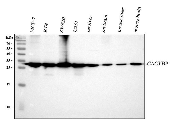
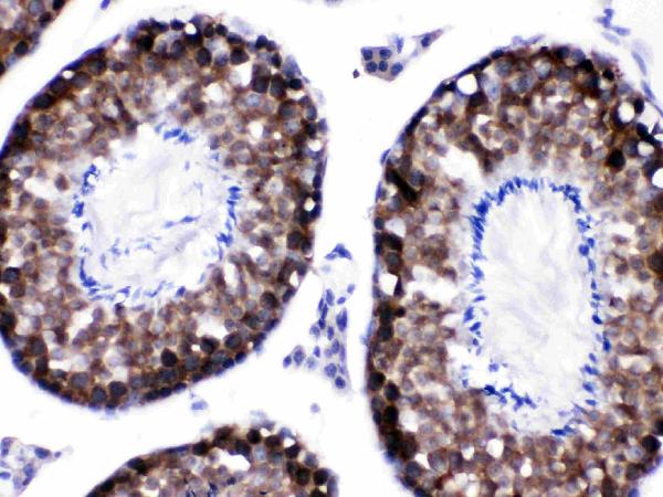
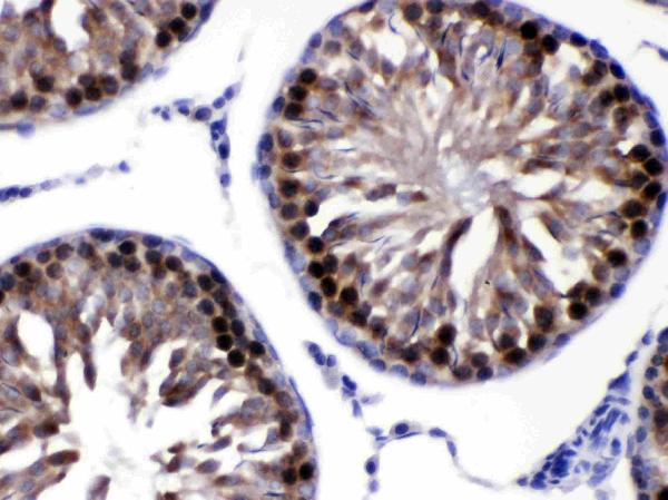
Facts about Calcyclin-binding protein.
Participates in the ubiquitin-mediated degradation of beta-catenin (CTNNB1) (By similarity). .
Mouse | |
---|---|
Gene Name: | Cacybp |
Uniprot: | Q9CXW3 |
Entrez: | 12301 |
Belongs to: |
---|
No superfamily |
CacyBP; calcyclin binding protein; GIG5; hCacyBP; MGC87971; RP1-102G20.6; S100A6-binding protein; S100A6BPgrowth-inhibiting gene 5 protein; Siah-interacting protein (SIP); Siah-interacting protein; SIPcalcyclin-binding protein
Mass (kDA):
26.51 kDA
Mouse | |
---|---|
Location: | 1|1 H2.1 |
Sequence: | 1; |
Highly expressed in brain and EAT cells. Expressed at low level in heart, muscle, and at very low level in the liver, spleen, lung, kidney and stomach.
The Boster Bio: Best Uses Of The Cacyp2C19 Antibody provides primary antibodies to the CACYBP Marker with high affinity. These antibodies have been validated for use in Western Blotting, Immunohistochemistry, and ELISA. They are well cited and trusted in the scientific community. We provide the highest quality antibodies for the best results. These antibodies are also produced in highly sensitive and specific concentras.
The primary antibody used with the CACYBP marker detects the expression of CacyBP/SIP. Although gastric tissues showed minimal expression of the protein, cancerous tissue expressed it in high levels. However, this study did not determine whether CacyBP/SIP expression is associated with other clinicopathological factors such as tumor size, TNM stage, and TNM resection. Further studies are needed to understand the role of CacyBP/SIP in the development of gastric cancer.
After incubation of the primary antibody with the marker, the membrane was washed three times with PBST buffer containing 0.5% Tween 20. Next, a horseradis peroxidase-conjugated anti-mouse IgG was added to the binding mixture. The results were analyzed by using a Tanon-410 automatic gel imaging system. The results are shown in a figure at the end of this article.
Knockdown of CacyBP inhibited viral replication and arrested apoptosis in DF-1 cells. The RNA-mediated knockdown of the CACYBP gene reduced the expression of the viral protein via multiple caspase-dependent pathways. The V protein, an inhibitor of viral replication, is involved in the regulation of CacyBP/SIP expression. The V protein binds to CacyBP and inhibits cell apoptosis.
The VC and VN antibodies showed similar results. The C-terminal domain of the V protein is essential for interaction with the CACYBP/SIP marker. The most overlapping fluorescence spots were observed in the cytoplasm. Further studies are necessary to validate the results of the co-immunoprecipitation experiments. Once these assays are complete, the researchers can perform quantitative analysis of CACYBP and V protein expression.
Using a secondary antibody with a specific antigen can be beneficial in several applications. These antibodies are often tagged with a reporter molecule, such as an enzyme like HRP or a fluorophore like FITC. The secondary antibodies can bind to the primary antibody, thereby increasing sensitivity and amplifying the signal. Secondary antibodies may be used alone or in combination with other primary antibodies.
One study found that CacyBP/SIP expression was associated with the progression of gastric cancer and a lower rate of tumor growth. It was also found that overexpression of this marker was associated with higher TNM stage and tumor differentiation. These findings suggest that the protein plays tumor-specific roles in cancer. However, further studies are needed to confirm the relationship between CacyBP/SIP and the various aspects of gastric cancer.
In a study that compared CacyBP/SIP expression levels in primary gastric cancer tissue samples, CacyBP/SIP expression was found in a high percentage of non-metastatic gastric cancer patients and 31% of metastatic patients. The results showed no significant difference between non-metastatic and metastatic gastric cancers. A future study will be necessary to confirm this finding. There are a variety of other ways to determine whether CacyBP is associated with gastric cancer and how to detect it.
CACYBP is a nuclear/cytoplasmic protein that participates in calcium-dependent ubiquitination. It has been associated with beta-catenin degradation. It interacts with S100 proteins such as SIAh2, SKP1A, and S100A6. It may be involved in calcium-dependent ubiquitination or act as a molecular bridge for the ubiquitin E3 complex. Two isoforms of the gene have been identified: human CacyBP/SIP and mouse SIP.
Depending on the downstream application of a secondary antibody, a number of label options may be available. In general, the secondary antibody will be labeled with the same antigen as the primary antibody. The secondary antibody should have a specific affinity to the target antigen to increase the detection sensitivity and to enhance the purity. Hence, the secondary antibody used with the CACYBP marker is critical in analyzing the CACYBP protein.
The most important thing to know when using the CACYBP as a marker for cardiovascular disease is the optimal time for incubation. Although there are many factors that determine the optimal time for incubation, a key factor is the presence of BIRC5 and SIP. They are proteins that interact with each other to form the Ubiquitin-ligase complex. These two proteins are important for the degradation of non-phosphorylated b-catenin.
Primary antibodies were purchased from DakoCytomation and diluted in a solution of Antibody Diluent (S 0809). Secondary antibodies were conjugated to horseradish peroxidase-labeled polymer. After incubation, sections were counter stained with QS haematoxylin. The sections were then examined under a light microscope.
Performing IHC/ICC experiments with a primary antibody requires optimization of all steps, including the concentration of the antigen to be detected. Optimal conditions are essential to detect specific staining, and the working dilution of polyclonal antibodies should be lower than that of monoclonal antibodies. A high quality antibody exhibits little or no cross-reactivity and a low working dilution is essential for the performance of your experiment.
The size of an Ig gene's combinatorial repertoire is huge, but its function is relatively limited. The combinatorial repertoire can be enormous, but its size is limited by the protective biological role of antibodies. In addition, antibody size is a crucial determinant of specificity. Luckily, this limit is well within the range of human antibodies. But how does one determine whether an antibody is specific? Here are some ways to determine the size of your antibody repertoire:
The sensitivity of a primary antibody is evaluated by comparing its OD450 value to that of a negative control. A negative control shows a high OD450 value, while a positive control shows a low OD450. The same test is done for antibodies to the same antigen. If a primary antibody is specific for a given molecular weight protein, it should label the same protein at the same molecular weight.
Another factor influencing the specificity of a primary antibody is its ability to bind a single antigen. Therefore, the antibody must be highly specific for the antigen that it is targeting. Absorption controls are not reliable, and their results may be false negative. Absorption tests are inaccurate if epitopes found on several antigens are shared, so an antibody may be able to bind to multiple antigens.
Controls for the specificity of primary antibodies include the use of a transfected cell line. The transfected cell line expresses the primary antibody antigen. The procedure includes fixing and stripping the cell in the same manner as the experimental sample. If the cells are not transfected with the desired protein, they serve as a negative control and have no antigen protein. In addition, siRNA is used to knock down the expression of the antigen protein.
PMID: 9572262 by Filipek A., et al. Molecular cloning and expression of a mouse brain cDNA encoding a novel protein target of calcyclin.
PMID: 10884380 by Nowotny M., et al. Characterization of the interaction of calcyclin (S100A6) and calcyclin-binding protein.