This website uses cookies to ensure you get the best experience on our website.
- Table of Contents
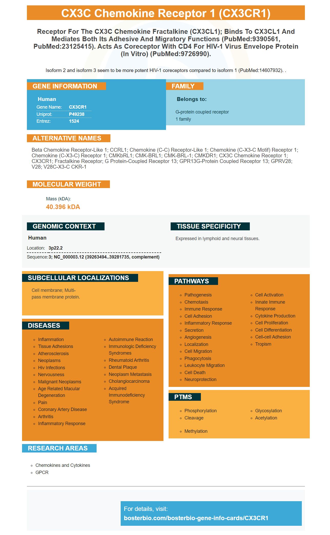
Facts about CX3C chemokine receptor 1.
Isoform 2 and isoform 3 seem to be more potent HIV-1 coreceptors compared to isoform 1 (PubMed:14607932). .
Human | |
---|---|
Gene Name: | CX3CR1 |
Uniprot: | P49238 |
Entrez: | 1524 |
Belongs to: |
---|
G-protein coupled receptor 1 family |
Beta chemokine receptor-like 1; CCRL1; chemokine (C-C) receptor-like 1; chemokine (C-X3-C motif) receptor 1; chemokine (C-X3-C) receptor 1; CMKbRL1; CMK-BRL1; CMK-BRL-1; CMKDR1; CX3C chemokine receptor 1; CX3CR1; Fractalkine receptor; G protein-coupled receptor 13; GPR13G-protein coupled receptor 13; GPRV28; V28; V28C-X3-C CKR-1
Mass (kDA):
40.396 kDA
Human | |
---|---|
Location: | 3p22.2 |
Sequence: | 3; NC_000003.12 (39263494..39281735, complement) |
Expressed in lymphoid and neural tissues.
Cell membrane; Multi-pass membrane protein.
You've come to a good place if CX3CR1 is a marker for your research. This article will explain Boster Bio and CX3CR1 siRNA lentiviruses. Boster Bio provides credits for results you share. Boster Bio offers discounts on CX3CR1 Lenviviruses.
Boster Bio has developed an ELISA kit for the detection of CX3CR1 protein. CX3CR1 is a chemokine molecule that recognizes ligands CD163, F4/80, ADAM17. This marker can be found both in human and mice tissues. Its levels in the peripheral blood can be determined by staining cells in the presence of a fluorescent reagent.
A Boster Bio CX3CR1 over expression lentivirus is available for use in the treatment of a variety of human cancers. It is designed to induce gene expression in cancer cells by transforming BMECs to express CX3CR1. These cells are more likely to recruit and multiply HCC cells in their spines if they have high levels of CX3CR1.
CX3CR1 was expressed in human cancer cells using an anti MMP13 antibody from Santa Cruz Biotechnology and an anti PTK2 antibody from Cell Signaling Technology. The Boster Bio CX3CR1 lentivirus was used for the transformation of MMP11b+/CD45+/Ly-6C.
The EFEMP1 protein acts as a suppressor to cell proliferation. The cells can't go from G1/G0 phase to S phase if it isn't knocked down. Over-expression of EFEMP1 promotes cell growth and increases the number of S-phase cells. The expression of EFEMP1 was also observed using Western blot, q-RT-PCR, and ICC.
The effects of DT upon tumor cells lasted three days. Microglia repopulation may not begin until seven days after the last DT injection. CX3CR1-overexpression lentivirus also prevented bone regrowth in mice, and human breast cancer cell lines. Researchers found that CX3CR1 expression was upregulated in spinal metastases. Additionally, CX3CR1 mRNA levels and protein expression levels were significantly higher in tumor bone than in normal vertebral.
Cancer cells also produce cytokines, which are substances that can be used to combat disease. These cancer cells can grow faster and contribute to the metastatic niche by increasing CX3CL1 expression. Boster Bio researchers used lentiviruses, which knocked out CX3CR1 from BMECs, in a previous study. These cells were then analyzed using CCK-8 as well as transwell assays to determine proliferation and migration.
Boster Bio scientists created a mouse model for cancer metastases using a nude mouse model. HCC cells were injected in the left ventricle of a mouse to test whether the CX3CR1 gene-over-expression lentivirus might increase tumor growth. After six weeks, mice underwent spinal metastases testing. Only two metastases could be observed in the control group. The CX3CR1MHCC97H mouse group had seven spinal metastases. The spinal metastases were also examined using micro-CT.
Boster Bio's lentiviruses target ICAM-1 (Immunoglobulin A) to promote transendothelial migration in NSCLC cell lines. The researchers used immunohistochemistry, western blotting, and reverse transcription-quantitative PCR to detect CX3CL1 and ICAM-1 expression. The lentiviruses significantly lowered ICAM-1 expression in VBMECs. These results suggest that CX3CL1 or ICAM-1 could be downstream effectors for CX3CL1 signaling.
The lentiviruses were tested in a variety of experiments. One of the experiments included a HCC mouse model and an animal model. Boster Bio sells an ELISA kit that can be used to detect CX3CL1 expression in vivo. After analyzing the results, the ELISA was used to determine the CX3CL1 levels in the cells.
GeneChem, Inc. provided plasmids to create the lentiviruses. The siADAM17 and CX3CR1 lentiviruses were used to transfect the cells. The manufacturer's instructions were followed during lentiviral transfer. To assess knockdown efficiency, we collected the cells 48-72 h after the transfection. The most efficient cells were chosen for the subsequent experiments.
In addition to BMECs, a mouse model of HCC metastases was established by injecting HCC cells into the left ventricle of the nude mouse. BMECs expressing CX3CR1 siRNA were transfected with an ADAM17 target siRNA. After six weeks, the mice were examined to determine if there had been spinal metastases. Mice who received tumor cells were found to have significantly larger tumors in CX3CR1MHCC97H than in the control group. The tumors were analysed by micro-CT.
NSCLC spinal metastases can be affected by the CX3CR1/ICAM-1 feedback loop. It also facilitates the extravasation of CTCs into vertebral cancellous bone, the first step of tumor spinal metastasis. The disease progression can effectively be stopped by inhibiting CX3CR1 within VBMECs. While this study was limited in mice, it provides important insights into the role of CX3CR1 in spinal metastasis.
Recent studies have shown that CX3CL1 plays a crucial role in the interaction of circulating NSCLC cells with VBMECs. In xenograft models, CX3CL1/ICAM-1 enhanced spinal metastasis. CX3CL1 also increased the permeability and adhesion of VBMECs, and tumor cell migration. Moreover, this inhibitor stimulated apoptosis and reduced tumor cell mortality in the experimental model.
PMID: 7590284 by Raport C.J., et al. The orphan G-protein-coupled receptor-encoding gene V28 is closely related to genes for chemokine receptors and is expressed in lymphoid and neural tissues.
PMID: 7646814 by Combadiere C., et al. Cloning, chromosomal localization, and RNA expression of a human beta chemokine receptor-like gene.