This website uses cookies to ensure you get the best experience on our website.
- Table of Contents
27 Citations 18 Q&As
9 Citations 16 Q&As
41 Citations 16 Q&As
7 Citations 15 Q&As
2 Citations
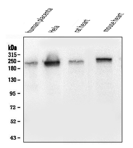
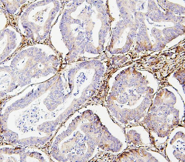
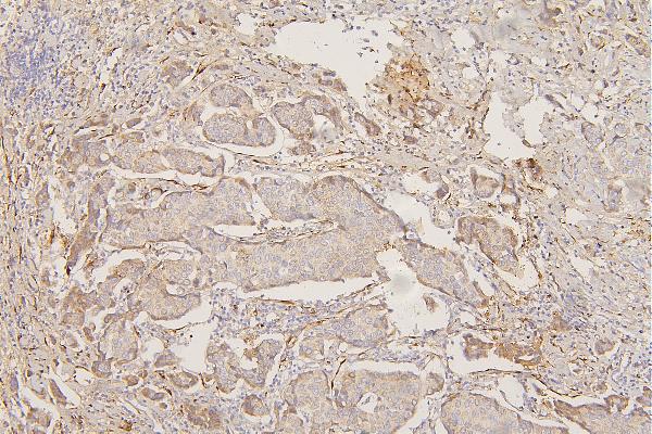
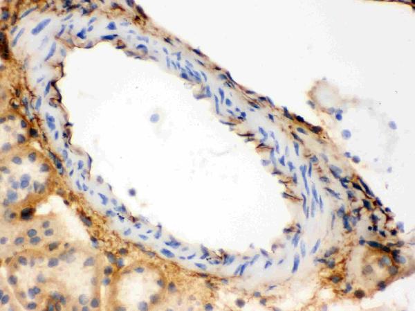
Facts about Collagen alpha-1(III) chain.
Is the significant ligand of ADGRG1 in the developing brain and binding to ADGRG1 inhibits neuronal migration and activates the RhoA pathway by coupling ADGRG1 to GNA13 and possibly GNA12. .
Human | |
---|---|
Gene Name: | COL3A1 |
Uniprot: | P02461 |
Entrez: | 1281 |
Belongs to: |
---|
fibrillar collagen family |
alpha1 (III) collagen; COL3A1; Collagen 3; collagen alpha-1(III) chain; Collagen III alpha 1; collagen, fetal; collagen, type III, alpha 1; Collagen-3; EDS4A; Ehlers-Danlos syndrome type IV, autosomal dominant; FLJ34534
Mass (kDA):
138.564 kDA
Human | |
---|---|
Location: | 2q32.2 |
Sequence: | 2; NC_000002.12 (188974373..189012746) |
Secreted, extracellular space, extracellular matrix.
This article will concentrate on the COL3A1 indicator, which is a type III collagen. Its role in coronary heart disease is explained. Learn about its interactions with and expression in the wall of the vascular system. This article will explore the numerous applications and limitations of this marker. Read on to learn more. COL3A1 is a type of collagen which is heavily expressed in the walls of the vascular wall.
The type III collagen gene encodes pro-alpha1 chains of the collagen type III protein. This type of collagen which is fibrillarin nature, is commonly found in connective tissue. This marker may also be related to chondrocyte hypertrophy. If it is analyzed using this method, it may determine the degree of collagen deposition in different tissues. It can be used to determine the causes of tissue degeneration.
The discovery of COL3A1 as a marker of coronary artery disease came as a shock. The genetic disorder was first identified by researchers studying Ehlers Danlos syndrome index patients of type IV. They could not identify any connection between specific mutations or types of complications. They identified 41 exon to intron Recombination mutations (EIR) and an ivs24+1G–A mutation in seven patients with index mutations and seven point mutations within the triple-helical region.
The researchers found an splice location mutation in the COL3A1 gene in a 40-year-old man with EDS. The patient didn't show any vascular anomalies, including a degeneration of vascular walls. The investigators found that the COL3A1 protein was found in a very low amount in the blood of the patient, who was homozygous for the COL3A1 mutation. Despite the presence of an inflammation disease in his arteries the patient's blood levels were normal.
In addition to the CCD and MDS, COL3A1 is also a risk factor for pregnancy-related morbidity. A study has shown that a G to A polymorphism COL3A1 creates a large Belgian family with EDS type IV. The phenotypes of the affected members of the Matton family was a mosaic for the AvaII polymorphism. Furthermore, the fibroblasts from the affected family secreted normal collagen. The fibroblasts also produced less type III collagen than cells that were used as control. Interestingly, the affected member of the Matton family had normal levels of mRNA of COL3A1 and a significant decrease in the type III collagen.
It is interesting to note that the COL3A1 gene is a genetic marker for coronary artery disease. It encodes a protein that is essential to the production of collagen, and a mutation in this gene can result in Ehlers-Danlos syndrome type IV. These mutations may increase the chance of developing coronary artery disease sixfold. It also affects the response of patients to antithrombotic therapy.
COL3A1 expression is present throughout the blood vessels. Mutations in COL3A1 can lead to different phenotypes similar to those found in the vascular form of EDS. In addition, IFN-gamma blocks the transcription rate of COL1A1 and COL3A1 genes, destabilizing their respective transcripts. Moreover, COL3A1 mutations lead to reduced procollagen chain production.
vEDS is caused by mutations in COL3A1 which is a genetic defect that affects collagen III's glycine rich domain. Symptoms of this disease are caused by autosomal dominant mutations in the COL3A1 gene, which encodes the pro-a1 chain of collagen III. The mutations resulting in abnormal protein formation are associated with more severe phenotypes and haploinsufficiency is associated with milder symptoms. Within the same family the severity and time of onset of disease can be very different. Other possible causes of the disease are unclear.
While vEDS is transmitted via autosomal dominant patterns, there have been reports of recessive inheritance. One family had two identical COL3A1 gene mutations that led to vascular-type EDS in a daughter. Numerous studies have suggested that vEDS is caused by genetic changes to the vascular wall. There have been changes to brain structure and function in cases of the vascular type of EDS.
Mutations in COL3A1 may cause premature death. Genetic changes that affect Col3A1 may lead to a shorter life span in mice. Type III collagen is created by the glycine mutations. It has lower thermal stability and is more prone to proteinases. Additionally, COL3A1 cells are enriched in COL3A1 proteins due to glycine mutagenesis. Ultrastructural analysis has demonstrated that collagen fiber diameters have changed.
A single-base mutation in COL3A1 results in an RNA splicing defect and a unique splicing pattern. Intron 37 is absent in the mutant allele, leading to an 80% splicing rate. At lower temperatures the abnormal splicing speed increased to 90% at 31 degrees Celsius. This is a new finding in platelet mutations.
This study shows that the endothelial NEDD9 protein mediates activated adhesion between platelets and PAEC by binding P-selectin. This study emphasizes the need to investigate the mechanisms that activate platelet activation in hypoxic conditions, particularly in PAH patients and PH patients. Patients with PAH/CTEPH must be considered hypoxic to better understand the role played by this protein in activating platelets.
The COL3A1 gene is found in multiple copies. The largest intron is 11.5 km2 and contains four copies of a 5-bp repeat. The smallest intron has eight bp. This implies that the COL3A1 gene is responsible for a wide range of functions. It is believed to be involved in the regulation of the function of platelets, which is vital for the development of healthy blood cells.
The COL3A1 gene provides instructions for type III collagen. Collagens are proteins that help strengthen different tissues in the body. Type III collagen can be found in the lungs, skin intestinal walls, the skin, and blood vessels. It interacts with platelets and plays an important role in blood clotting. COL3A1 mutations can trigger vascular Ehlers Danlos syndrome the condition in which people are deficient in type III collagen.
Cell Reports published an article that suggests that NEDD9 regulates the COL3A1 nerve node, a major one in the transition between adaptive and pathogenic fibrosis. This finding could have therapeutic implications, as the regulation of COL3A1 may help prevent vascular remodeling in patients with PAH. NEDD9 also regulates the production and release of collagen. The study also showed that NEDD9 blocks several genes involved in vascular remodeling, including COL3A1.
The role of NEDD9 was identified by identifying the amino acids that are specific to a particular residue, Cys18, in the Src homology domain (SH) of NEDD9. The results of molecular simulations revealed that oxidative modification caused by NEDD9 blocks its binding to SMAD3, a protein implicated in pulmonary artery fibrosis. Although the mechanism behind NEDD9 modulation isn't fully comprehended, these findings demonstrate that NEDD9 is a critical participant in the vascular remodeling of the lung.
In the study, we observed that si-NEDD9 decreased the levels of Cys18 and NKX2-5 in HPAECs. We also found that si NEDD9 reduced the expression of SMAD3, and ALDO increased the binding of NEDD9 by COL3A1. It is interesting that despite the inhibition of NEDD9, it didn't affect the NKX2-5-mediated COL3A1 transcription.
The study also showed that NEDD9 overexpression correlates with higher levels of collagen III, 3D collagen matrigel contraction and cell stiffness. Further, ALDO induces several pro-fibrotic changes in HPAECs, including the formation of distinct "net" pattern of collagen stranding that is not apparent in normal human lung fibroblasts.
PMID: 2764886 by Ala-Kokko L., et al. Structure of cDNA clones coding for the entire prepro alpha 1 (III) chain of human type III procollagen. Differences in protein structure from type I procollagen and conservation of codon preferences.
PMID: 11566270 by Valkkila M., et al. Genomic organization of the human COL3A1 and COL5A2 genes: COL5A2 has evolved differently than the other minor fibrillar collagen genes.
*Showing only the more recent 20. More publications can be found for each product on its corresponding product page