This website uses cookies to ensure you get the best experience on our website.
- Table of Contents
Facts about Circadian locomoter output cycles protein kaput.
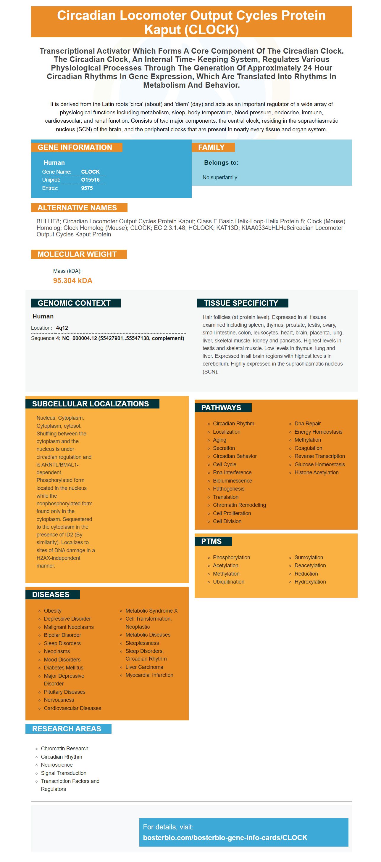
It is derived from the Latin roots 'circa' (about) and 'diem' (day) and acts as an important regulator of a wide array of physiological functions including metabolism, sleep, body temperature, blood pressure, endocrine, immune, cardiovascular, and renal function. Consists of two major components: the central clock, residing in the suprachiasmatic nucleus (SCN) of the brain, and the peripheral clocks that are present in nearly every tissue and organ system.
Human | |
---|---|
Gene Name: | CLOCK |
Uniprot: | O15516 |
Entrez: | 9575 |
Belongs to: |
---|
No superfamily |
BHLHE8; circadian locomoter output cycles protein kaput; Class E basic helix-loop-helix protein 8; clock (mouse) homolog; clock homolog (mouse); CLOCK; EC 2.3.1.48; hCLOCK; KAT13D; KIAA0334bHLHe8circadian locomoter output cycles kaput protein
Mass (kDA):
95.304 kDA
Human | |
---|---|
Location: | 4q12 |
Sequence: | 4; NC_000004.12 (55427901..55547138, complement) |
Hair follicles (at protein level). Expressed in all tissues examined including spleen, thymus, prostate, testis, ovary, small intestine, colon, leukocytes, heart, brain, placenta, lung, liver, skeletal muscle, kidney and pancreas. Highest levels in testis and skeletal muscle. Low levels in thymus, lung and liver. Expressed in all brain regions with highest levels in cerebellum. Highly expressed in the suprachiasmatic nucleus (SCN).
Nucleus. Cytoplasm. Cytoplasm, cytosol. Shuffling between the cytoplasm and the nucleus is under circadian regulation and is ARNTL/BMAL1-dependent. Phosphorylated form located in the nucleus while the nonphosphorylated form found only in the cytoplasm. Sequestered to the cytoplasm in the presence of ID2 (By similarity). Localizes to sites of DNA damage in a H2AX-independent manner.
When it comes to Boster Bio's CLOCK Marker Researchers often are wondering what the best uses of the product are. In this article, you'll find the three most popular uses for the Boster Bio anti-Macrosialin CD68 antibody as well as the CLOCK Marker. Its primary function is to detect epithelial markers.
The Boster Bio Anti-Macrosialn CD68 Monoclonal (MacM68) Antibody has been tested in IHC and WB applications. It reacts with Human, Mouse, and Rat. The product contains 0.05% Thimerosal. The Boster Bio Anti-Macrosialin CD68 Antibody has no hazard and is suitable for production in-house.
The antibody recognizes CD68 which is a protein that is highly expressed in a wide range of cells including macrophages, mononuclear and phagocytes. This protein is also detected by immunohistochemistry. It is highly expressed in macrophages in tissues, dendritic cells and is a crucial marker to determine the macrophage lineage. It can be used to differentiate similar-looking diseases.
Macrosialin is a transmembrane glycoprotein that is expressed on the surface of human macrophages. The anti-Macrosialin CD68 antibody is an excellent instrument for biological assays, and Boster Bio uses rabbit and mouse blood samples to formulate its Reagents. While its exact function is not yet fully known, it is believed to play an important role in phagocytic cell and the intracellular lysosomal system. Moreover, it may play an important role in extracellular cells-cell interactions with substrates.
To use Boster Bio Anti-Macrosial-CD68 Antibody, be sure to adhere to the suggested storage conditions. The product should be stored at 4 degrees Celsius or lower and should not undergo repeated freezing and freezing and. The shelf duration of this anti-Macrosialin antibody is 18 months. If you do not plan to use the product during this timeframe, you may need to consider purchasing it on dry ice.
Its binding affinity to specific tissue lectins makes it particularly effective in lung cancer. Macrophages typically are characterized by underlying subsets of cells, and the anti-CD68 antibody will target the cells that are able to harbor these underlying subsets. This anti-MacrosialinCD68 antibody will not be clinically effective due to the diversity of macrophage populations.
The boster Bio Anti-Macrosialin-CD68 Antibody recognizes CD68 in a range of cell types, including monocytes and granulocytes. It also plays an important role in macrophage recruitment and activation. It is a member of the Lysosome associated Membrane Protein (LAMP). The anti-Macrosialin CD68 antibody aims to block the cytotoxic effects of phagocytosis and enhance the immune system by enhancing its function.
Boster Bio's monoclonal antibodies against keratin were created. They are able to detect and stain a broad variety of cytokeratins derived from epithelial tissues that are malignant and normal. This monoclonal antibody is able to demonstrate high specificity to detect epithelial cells in a variety of human tumors.
The antibody solution can be stored for up to 24 months. Boster Bio encourages scientists and researchers to submit results from their own research. They may be eligible for credit for product. The reagents can only be used for research purposes, and not for diagnostic or drug applications. It is also appropriate for all scientists around the globe. This product is available in various locations.
Antibodies against the cytokeratin (CK7) were used to establish human bronchial epithelial bronchial cell cultures. Anti-CK7 antibodies help distinguish epithelial cells and fibroblasts. Antibodies against the types I, III and IV collagen strongly reacted with epithelial cells but did not affect fibroblasts. Epithelial cells did not react strongly to antibodies to fibronectin. However, they did not react with the antibody against the fibronectin.
The study's results revealed that OV-TL12/30 was present in most superficial conjunctival epithelial cells. Contrasts of other tested antibodies revealed that less than 25 percent epithelial cells are present on the cornea. The antibody was an accurate marker for LSCD. Positive tests are found in 78 percent of patients.
The antibody proved to be highly effective in detection of CK7 by RT-PCR. GAPDH was employed as an internal control to determine whether CK7 expression was present in the corneal epithelium of the central cornea. Positive cells appeared first in the limbal epithelial layer . They then increased in the conjunctival epithelium. Table 2 lists the percentage of cells that are positive for CK7 and the presence of GCs.
PMID: 10198158 by Steeves T.D.L., et al. Molecular cloning and characterization of the human CLOCK gene: expression in the suprachiasmatic nuclei.
PMID: 11441146 by Rutter J., et al. Regulation of clock and NPAS2 DNA binding by the redox state of NAD cofactors.