This website uses cookies to ensure you get the best experience on our website.
- Table of Contents
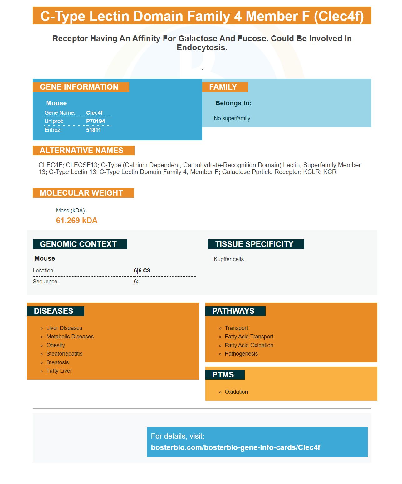

Facts about C-type lectin domain family 4 member F.
.
| Mouse | |
|---|---|
| Gene Name: | Clec4f |
| Uniprot: | P70194 |
| Entrez: | 51811 |

| Belongs to: |
|---|
| No superfamily |

CLEC4F; CLECSF13; C-type (calcium dependent, carbohydrate-recognition domain) lectin, superfamily member 13; C-type lectin 13; C-type lectin domain family 4, member F; Galactose Particle Receptor; KCLR; KCR
Mass (kDA):
61.269 kDA

| Mouse | |
|---|---|
| Location: | 6|6 C3 |
| Sequence: | 6; |
Kupffer cells.


This article will introduce you the CLEC4F macrophage marker, a local liver marker. We will discuss how it may be involved in Listeria moncytogenes infection, modulate glycolipids presentation, or how it could be a potential target for nanobodies. We will also talk about how CLEC4F can be useful in research on immunomodulation.
CLEC4F is an extremely specific protein found in resident macrophages of the liver. It is unknown exactly how this protein functions in the liver. It is not clear what its role is in the liver. However it may be involved in the regulation or glycolipid antigenic presentation in Kupffer cells and in the removal of desialylated platlets. This protein has been identified by KCs as an important marker.
The liver has the highest number of macrophages within the mammalian body. These cells are vital participants in various liver processes. Acute hepatotoxicity can cause blood monocytes to colonize liver. These monocytes can also colonize bone marrow. Although the function and functions of resident liver macrophages are not known, it is believed they have distinct functions.
Although KCs are not directly involved in the immune system in the liver, they have been implicated in many diseases. Nonalcoholic fatty liver disorder (NAFLD), steatosis, and inflammation are both inflammatory diseases that increase the risk for mortality and insulin resistance. Both conditions are caused in part by abnormalities of KCs. A Nature study that examined the role Clec4F played in liver disease revealed that the protein was a key mediator in NAFLD.
CLEC4F was only detected in freshly isolated Kupffer cells. It was not detected in bone or peripheral blood leukocytes. It is expressed as polypeptides with varying molecular weights in the presence tunicamycin. CLEC4F was detected in an incompletely glycosylated form 70 kDa in a study with 293 T cells.
The CLEC4F C lectin, which is a member of C-type lectins superfamily, has varied binding specificities. It can be expressed on Kupffer lectin cells and infiltrating monkey cells in the liver. It could play an important role modulating the presentation and distribution of glycolipids in Kupffer cells. It is also known for its ability to influence the activity NK-cells, which express CD1d, restriced ab TCRs.
To test this hypothesis, researchers used anti-CLEC4F monoclonal antibodies and a mouse model lacking CLEC4F expression. CLEC4F is co-expressed on Kupffer cellular cultures in mice subjected to L. monocytogenes. CLEC4F was found to be expressed on liver Kupffer cells as well as F4/80-positive Kupffer cell lines. It is not expressed on peritoneal macrophages and other macrophage cell line. They found that CLEC4F-/ mice were not affected by L. monocytogenes infection. Both antibodies were used to confirm the presence CLEC4F in liver tissue, as is the case with the Boster bio CLEC4F-/ mouse models.
Only the livers of wild type and mutant mice contained the CLEC4F protein. It is not detected in peripheral blood leukocytes that contain monocytes. The anti-CLEC4F antibacterial antibody detected polypeptides with molecularweights of 100 to 61kDa. Anti-CLEC4FMAb decreased CLEC4F molecularweight to 61 kDa using 293 T cell.
CLEC4F is also found in embryogenesis in different tissues. This protein regulates the microenvironment in the liver. It can also be found in the yolk-sac. During embryogenesis, CLEC4F is detected in liver, yolk sac, and embryo. It may have a role in modulating the presentation of glycolipids. But, further studies are needed to confirm the role of this protein in the liver.
The immune response to Listeria monocytogenes infections is mediated by the CLEC4F marker. It is caused by L. monocytogenes-infected inflammatory monocytes. The liver is the main organ responsible for clearing blood-borne bacteria. Infection with this pathogen could lead to the recruitment of inflammatory monocytes. CLEC4F expression decreased in mice after being exposed to Alk1fl/fl/fl vav1Cre mice.
CLEC4F belongs to the C-type lectin familia and has a high binding specificity for glycolipids. CLEC4F expression is found in Kupffer cells in liver and infiltrating monkeys. Its function in infection remains unclear. The exact role of CLEC4F is still unknown.
CLEC4F, an a-GalCer antagonist, is expressed only on resident Kupffer cells, but not on infiltrating F4/80+ monoocytes. Further research is needed to determine if CLEC4F plays a role in immune modulation and the development of antibiotic resistant. It is crucial to understand how the CLEC4F-receptor functions in Listeria monocytogenes disease.
ALK1 is necessary for KC-mediated Listeria monocytogenes capture. A lack of ALK1 causes failure to capture the bacterium and consequently, overwhelming disseminated infection. In addition to inhibiting bacterial virulence, ALK1 also provides an important signal for KC-mediated Listeria monocytogenes infection.
CLEC4F and F4/80 are co-expressed on Kupffer cells in the liver. Parafilm-embedded liver slices were used for immunohistochemistry. The nuclei were counterstained with Hoechst 33342. Confocal microscopy confirmed that CLEC4F and FL4/80 coexpression was present. The peritoneum was unaffected by the expression of F4/80 or CLEC4F. In addition to CLEC4F, F4/80-specific antibodies were used as isotype controls.
The Clec4F gene is a potential target of nanobodies. We created nanobodies which target Clec4F through inserting appropriate gene sequences to the pHEN6c plasmid. This was then transfected into E. coli HK6 host cells. After purification the nanobodies had to be extracted by osmotic stimulation and were then expressed using 400 mL culture media. Purification was accomplished by immobilized metallic affinity chromatography using an Ni-NTA columns. The nanobodies were stained using Coomassie Brilliant Blue staining, and SDS -PAGE.
We tested 99mTcClec4F nanobodies that were bound to recombinant Clec4F in experiments. These nanobodies then were tested in naive mice.
By cloning VHH from B-cells, we created a library Clec4F-specific nubodies. After selection by biopanning (phage-display), we discovered that Clec4F's specific nanobodies contained a great diversity. Based on their sequence identity within CDR3, which is the region of VHH where they vary the most, we divided them into 14 groups.
CLEC4F is another reason to target nanobodies with the antibody. It is highly specific for CD8+ cells and can be used for single-photon emission computed tmography and positron emission tomography. The anti CD11b antibody, also known c-FLAG (or c-FLAG), was used to detect inflammation and spleen disease in a mouse model.
Nanobodies can be used as imaging agents to treat cancer. Because of their high affinity and specificity, nanobodies are ideal imaging agents. Additionally to imaging, nanobodies may also be used in diagnostics and therapeutics. However, imaging may require special labeling. However, nanobodies are a promising option for cancer research. They can offer both diagnostic and therapeutic benefits in the fight to cancer.
CLEC4F is a protein expressed from peripheral blood lymphocyte cells. The total mRNA was extracted and converted to cDNA for use with phage enrichment ELISA. Antigen-specific antigen phages were enhanced by a phage enhancement ELISA using a third panning step. These phages could then be used for DNA sequencing.
CLEC4F type II transmembrane protein that is part C-type, lectin superfamily. It binds galactose and N-acetylgalactosamine. In the mouse, CLEC4F forms a coiled-coil interface and comprises conserved carbohydrate-recognition domains. CLEC4F shows distinct distances between its Ca2+ binding and its carbohydrate binding sites.
The CLEC4F marker in KCs is a useful tool. It is found in many tissues, including the yolk sac and fetal liver. Listeria monocytogenes infection is indicated by its presence in liver abscesses. It is found in infiltrating mononuclear cells surrounding liver abscesses. It is infected with Listeria monocytogenes in the liver and is strongly bound by Gal. Fucosylation inhibits it, but Globo H does not.
CLEC4F was coated onto 96 well plates with 1 mg/mL. This is the most common assay. It was left at 4°C overnight. PBS containing 4% skimmedmilk blocked the protein binding site. Clec4F nanobodies were then mixed in 1% skimmed PBST, and flown over a 400-RU recombinant mouse Clec4F protein coupled with a CM5chip. This assay confirmed that Ly6C and Clec4F were highly correlated.
PMID: 16141072 by Carninci P., et al. The transcriptional landscape of the mammalian genome.
PMID: 21183079 by Huttlin E.L., et al. A tissue-specific atlas of mouse protein phosphorylation and expression.