This website uses cookies to ensure you get the best experience on our website.
- Table of Contents
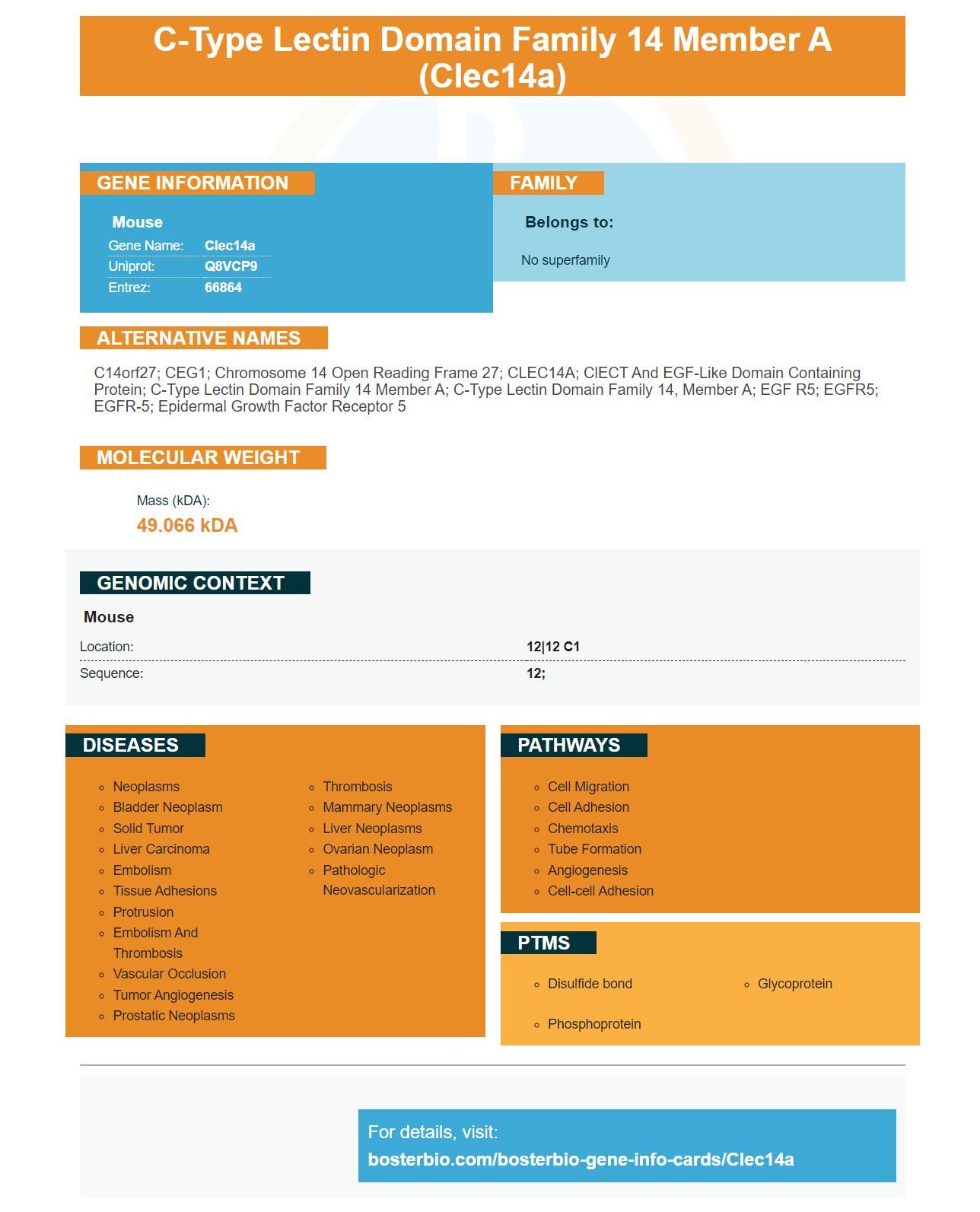
Facts about C-type lectin domain family 14 member A.
Mouse | |
---|---|
Gene Name: | Clec14a |
Uniprot: | Q8VCP9 |
Entrez: | 66864 |
Belongs to: |
---|
No superfamily |
C14orf27; CEG1; chromosome 14 open reading frame 27; CLEC14A; ClECT and EGF-like domain containing protein; C-type lectin domain family 14 member A; C-type lectin domain family 14, member A; EGF R5; EGFR5; EGFR-5; Epidermal growth factor receptor 5
Mass (kDA):
49.066 kDA
Mouse | |
---|---|
Location: | 12|12 C1 |
Sequence: | 12; |
Although CLEC14A/PECAM1 levels can vary among samples of the same patient's blood, they are relatively stable when compared to different tissues from the same patient. They can be used to analyse a wider range tissue types. This allows for more reliable measurements of tissue differences. Boster Bio's website has more information. Read the complete article: The Best Uses of the CLEC14A Symbol
We found that tumours lacking CLEC14A had higher levels of microvessels in the area and CD31-positive regions. The results suggest that CLEC14A might be involved in EC growth and angiogenic blood vessels integrity. This study also revealed a new mechanism that regulates tumour angiogenesis in response VEGF-A. We hope to understand how this mechanism works and what its implications are for treatment.
The role of CLEC14A in angiogenesis is not entirely understood. It is known, however, that the protein regulates VEGF C/VEGFR-2 pathways. This has been implicated in the progression and development of neovascular conditions. It may also play a part in lymphangiogenesis. Therefore, drugs that target this protein could be a promising treatment. More research is needed to fully understand the function and role of CLEC14A.
CLEC14A plays a role in regulation of VEGFR-2 expression and VEGFR-3 expression. Loss of CLEC14A reduces VEGFR-3 expression, and a lack of CLEC14A promotes VEGFR-2 expression. Therefore, tumor growth and progression are affected by the absence of CLEC14A. It increases VEGFR-2 and VEGFR-3 expression.
CLEC14A and a variety of other markers are expressed by ECs from both species. The expression of cell adhesion molecules, cell transcription factors (ERG), and Tbx2 help to identify subpopulations. The angiopoietin inhibitor TIE1 is also expressed by general endothelial tissue cells.
A-Smooth muscle actin was less abundant in tumours with reduced CLEC14A expression. Collagen type IV was also less abundant. The loss of these proteins will determine whether tumor angiogenesis promotes growth or suppresses it. In addition, mice with CLEC14A-KO tumors exhibited significantly lower levels of a-Smooth muscle actin coverage in CD31-positive tumours. CLEC14AKO cancers also had less collagen type 4.
These ECs not only express CLEC14A but also genes that are involved gas exchange and immune cells homing. They also contain genes for nuclear, membrane, secreted, and venous proteins. These ECs are involved diapedesis. Tumour ECs are also known to express CD248, CD93, and have been found to closely resemble each other. Thrombocytes also possess a number of genes including Akt, PDE7B, and others.
In a previous study the concentrations in human fibroblasts of CLEC14A (PECAM1) and CLEC14A (CLEC14A) were different. These levels were identical in glioma, adipose and human fibroblasts, suggesting that both proteins play an essential role in vascular function. Thus, a higher concentration of PECAM1 is desirable for treating fibrosis.
CLEC14A is expressed in myogenic cells, which are not necessarily derived from adipose tissue. These cells are located in a satellite cells niche and don't express any markers of endothelial or other cells. This suggests CLEC14A overlaps well with other cell populations. However, it creates a new population in human muscles. Our data suggest that CLEC14A could play a role in the pathogenesis and development of cancer cells.
The role of PECAM1 in regulating endothelial cell permeability and leukocyte trafficking has been shown in animal studies. It also functions as a stress response protein, increasing endothelial growth and improving blood permeability. Our results also suggest that PECAM1 is critical for maintaining the integrity of the vascular permeability barrier.
This study also showed that human muscle fibres made from cells lacking PAX7 are capable of regenerating when transplanted into an ectopic TA mouse. PAX7neg cell transplants form muscle fibers at the same rate as human muscle fibers. Induction of PAX7 caused molecular profiling changes in PAX7null cells. Further, PAX7 also induced MYOD1 while downregulating CLEC14A. These findings suggest that PAX7 may be a potential stem cell to regenerate human muscle fibers.
The CLEC14A proteins and gene are found in human tumors and healthy tissues. To develop new cytotoxic immunotherapies, it is essential to understand how these proteins are expressed. It is unclear how this marker can be used for clinical applications. To date, the CLEC14A gene and protein are being used for research and clinical applications. This article summarizes the current state of the CLEC14A protein and its future direction.
CLEC14A expression levels were low in healthy tissues suggesting that it was not associated for tumour growth. In tumour samples, CLEC14A mRNA levels were high, and high ratios between CLEC14A and TIE1 indicated that this gene is expressed more frequently in cancer cells. This finding supports the idea that other mechanisms may contribute to the expression of CLEC14A. The activin-receptor-like kinase 1 that is a member TGF-beta's superfamily regulates the expression of the protein.
Immunostaining with the CD31 antibody revealed that the CLEC14A protein was found in lung tissues. This antibody is used to identify endothelial cell types. CLEC14A was absent in the majority of tissues from healthy animals. However, it was found in cancerous tissues at low levels. The protein was detected within the alveoli of all lung specimens, two of fiveplacental tissues, one small intestine and one of five placental tissues. CLEC14A staining, although low in tumour tissue, was detectable in all organs.
The CLEC14A target is an immunotherapy target based on the level of C lectins expressed in tumor tissues. CLEC14A promotes angiogenic activity, but it also plays important roles during endothelial cell proliferation and tube formation. It is controlled by shear stress and elevated in solid tumours. CLEC14A is a positive marker in more than half the cases of renal cell carcinoma.
The CLEC14A anti-mouse antibody was developed by using the Noy and colleagues' method. It was used with an ImmPRESS Excel AMPLIFIZED HRP Polymer Staining Mouse IgG kit, purchased from Vector Laboratories. This antibody is extremely specific and reduces tumour growth. It is being evaluated for clinical applications.
The toxicity is a key question in evaluating the efficacy and safety of CLEC14A-targeted treatment. Some reports report reduced toxicity. However, there is a lot of evidence that this treatment doesn't cause serious side effects. It actually has several benefits. The first benefit of targeting CLEC14A is that it can be used in combination with other treatments.
The expression pattern CLEC14A of tumor cells and surrounding tissues has a large impact on the potential for toxicities. Some antigens that are thought to be tumour-causing agents have been shown, for instance, to cause severe toxicities within healthy tissues. This makes it important to characterize the expression of tumour antigens in healthy tissues to determine the potential off-target toxicity. This is important not only for predicting possible toxicities, but also to help select patients who will benefit from the therapy.
Tumours are not the only thing that can be caused by CLEC14A. These tissues include gastric cancer, alcoholic hepatitis, interstitial lung disease, and lung idiopathic fibrosis. To determine the toxicity of these therapies, further research is needed on the expression patterns CLEC14A within these tissues. This therapy is a promising option in the development of anti-cancer therapies.
Furthermore, the safety of drugs is a major factor in the efficacy or failure of CLEC14A targeted therapy. Although it is possible that these treatments can cause severe toxicity, there are still some promising options. For instance, aCLEC14A-targeted antibody may inhibit tumor angiogenesis by preventing cell-to-cell contact. However, these drugs can also cause an increase of cancer cell proliferation.
Recent research revealed that CLEC14A gene expression in the liver and lung is highly dependent on the blood flow. This is why further research is required to confirm the efficacy and safety of these drugs. Human patients have not been tested for the toxicity of CLEC14A -targeted therapies. Recent research has shown that the treatment did in fact not cause any toxicities in mice.
PMID: 16141072 by Carninci P., et al. The transcriptional landscape of the mammalian genome.
PMID: 21183079 by Huttlin E.L., et al. A tissue-specific atlas of mouse protein phosphorylation and expression.