This website uses cookies to ensure you get the best experience on our website.
- Table of Contents
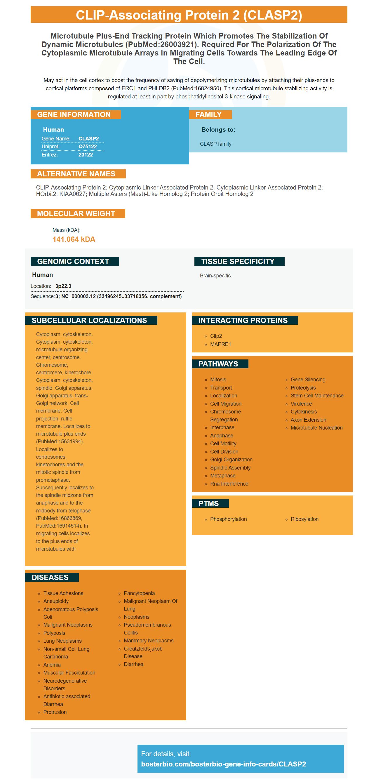
Facts about CLIP-associating protein 2.
May act in the cell cortex to boost the frequency of saving of depolymerizing microtubules by attaching their plus-ends to cortical platforms composed of ERC1 and PHLDB2 (PubMed:16824950). This cortical microtubule stabilizing activity is regulated at least in part by phosphatidylinositol 3-kinase signaling.
Human | |
---|---|
Gene Name: | CLASP2 |
Uniprot: | O75122 |
Entrez: | 23122 |
Belongs to: |
---|
CLASP family |
CLIP-associating protein 2; cytoplasmic linker associated protein 2; Cytoplasmic linker-associated protein 2; hOrbit2; KIAA0627; multiple asters (Mast)-like homolog 2; Protein Orbit homolog 2
Mass (kDA):
141.064 kDA
Human | |
---|---|
Location: | 3p22.3 |
Sequence: | 3; NC_000003.12 (33496245..33718356, complement) |
Brain-specific.
Cytoplasm, cytoskeleton. Cytoplasm, cytoskeleton, microtubule organizing center, centrosome. Chromosome, centromere, kinetochore. Cytoplasm, cytoskeleton, spindle. Golgi apparatus. Golgi apparatus, trans-Golgi network. Cell membrane. Cell projection, ruffle membrane. Localizes to microtubule plus ends (PubMed:15631994). Localizes to centrosomes, kinetochores and the mitotic spindle from prometaphase. Subsequently localizes to the spindle midzone from anaphase and to the midbody from telophase (PubMed:16866869, PubMed:16914514). In migrating cells localizes to the plus ends of microtubules with
In this article we will discuss how to use CLASP2 to increase cell proliferation in the CCK-8 experiment. We'll also show you how to perform Immunoblotting on mice and rats with anti-E-cadherin, and anti-vimentin antibody antibodies. We'll also be discussing the complexities and best uses of the CLASP2 marker.
Boster Bio Cell Proliferation Assasin CCK-8 is a rapid and easy method to measure cell proliferation rates in culture. It contains a Tetrazolium salt, which receives electrons of viable cells and transfers them into the dilution CCK-8 in the culture media. CCK-8 cannot be used for cell staining because it is highly water-soluble. However, it does retain its stability at -20degC and is stable for 2 years.
Cell Counting Kit-8 is a highly water-soluble tetrazolium sodium salt, WST-8. This makes it an ideal solution for sensitive colorimetric cell proliferation tests. When reduced, WST-8 gives off a yellow-colored formazan dye, which is soluble in tissue culture media. The WST-8 cell proliferation assay provides accurate results and is easy to use. The BosterBioCCK-8 is an excellent choice to work with live cells within the laboratory.
Boster Bio CCK-8 cell proliferation assassassassassassassassassassassa's uses a single-bottle solution that allows for fast and accurate cell counting. It is stable at 4 degrees Celsius because it is not radioactive. It doesn't require any premixing. The BosterBio CCK-8 cell proliferation assassassassassassassassassassassay is fast, precise, and easy-to-use
Boster Bio's CCK-8 cell proliferation assay assay has a low baseline level. This means that the assay has a higher sensitivity than other tetrazolium sodium salts. Furthermore, the Boster CCK-8 enzyme assay is highly sensitive. Its high sensitivity allows it to detect small quantities of DNA and RNA from samples of a wide variety of species.
When designing experiments, you should consider the frequency of the target cell. It is important to know how many cells each experiment requires. This will prevent you from repeating the exact same experiments. Table 1 shows how many cells are found in common rat tissues. Visit the dedicated page on cell frequency for more information. Table 2 lists the frequency of markers that are specific for immune cell lineages. There are dedicated antibodies for each cell type.
The immune system functions can be attributed to CD4+ T-cells. To detect this cell surface antibody, we used the mouse-anti-rat CD11b antibody. The CD106 anti mouse rat CD106 antibody recognizes CLASP2Marker, which is a 110kDa inducible transmembrane glyprotein. These samples were analyzed with FlowJo software. We also stain dead cells with Hoechst 33342.
To determine the expression CLASP2, peripheral samples of blood were collected from each animal using retroorbital vasculature plexus. To collect blood samples, EDTA -coated tubes were used. Blood counts were done using a Vet'ABC counting machine. Serum erythropoietin levels could be determined using ELISA or Quantikine mouse/rat EPO immunoassay. All samples were measured in duplicate using an internal quantity standard.
The murine CLASP2 protein gene spans 200kb and includes 40 exons. The second exon, common to all isoforms of CLASP2, was interrupted by a EGFP-loxP-pMC1NEO-loxP cassette. The target vector includes a neomycin resistance and a thymidinekinase gene to counter selection.
Immunoblotting rats and mice with Clasp2 marker showed that CLASP2 protein levels are similar throughout the hematopoietic tissue. However, association with the cell cortex requires the C terminus to be expressed. CLASP1 is expressed in mice at lower levels than CLASP2.
Bio-Rad produces a large range of anti-CLASP2 antibodies for flow cytometry, western blotting, and immunoprecipitation. Their antibodies can be used in flow cytometry. They can be used in flow cytometry by being conjugated with fluorescent dyes. The company also provides pre-prepared 2-, 3-, and 4-color cocktail reagents and mouse isotype controls.
The primary antibodies used in this study were goat anti-E-cadherin (#E-Cadh), anti-b-catenin (#BCAT), and anti-E-cadherin from the Boster Bio catalog. After 10 minutes of permeation with 0.3% Triton X-100, the cells were fixed in 2-4% Methane and then blocked with 3% sheep serum. The cells were then incubated at room temperatures with the primary antibodies three times with PBS. Next, they were probed for specific target proteins.
Primary antibodies used in immunoblotting the human tissues were abcam monoclonal b–Catenin (Dako monoclonal m3612), Boster polyclonal BB1771 (Molecular Devices' anti–Ki67 antibody) and Dako monoclonal m3612. Secondary antibodies were used in the detection of E-cadherin (LRRC4), Vimentin (LC3B), and AKT. After staining, signals were visualized with an Ultra SensitiveTM SP system and peroxidase detect.
E. coli expressing E-cadherin. This immunogen was used in immunoblotting to detect E-cadherin in the cells. Anti-Ecadherin can be used to identify the main components of a cell's cell's cytoplasm or membrane. It is also a good marker of its architecture. It helps researchers understand cell-cell adhesion junction functions.
E-cadherin might play an important role when tumors develop and is involved with mechanisms that regulate epithelial differentiate. Its loss has been linked with tumor invasiveness. It is an extracellular calcium binding protein that cleaves when Ca2+ is present. E-cadherin is a sensitive target to trypsin when it is cleaved.
The overexpression of LRRC4 in CRC cells inhibits collective cell invasion via a PI3K/AKT pathway. The knockdown of LRRC4 by Ube2v1 cells reduced expression of Ecadherin in CRC cells. Furthermore, knockdown of Ube2v1 rescued the down-regulation of p110a and LC3-II in Ube2v1-deficient cells.
The Simple WesternTM method detects human and mouse E-Cadherin using mouse embryonic carcinoma cell lines. It can also detect human epithelial and breast cancer cells using a goat anti–goat secondary antibody. The Simple WesternTM method can identify the E-Cadherin proteins at a 128 kDa molecular mass. This protein is blotted in reduced conditions using a 12-230kDa separation method.
An immunoblotting procedure using an antivimentin antibody on mice is a common way of determining if a protein is present in a particular type of cell. The protein is found in mesenchymal tissues and is responsible of controlling cell shape, mobility, and resistance to mechanical stress. Monoclonal mice anti-vimentin antibody reacts specifically with vimentin. The antibody can be used to identify vimentin in a variety of tissue samples.
The antibody LOB7 recognizes coil 2 of vimentin. The specificity of the antibody LOB7 was further confirmed by mass spectrometry. This antibody detects protein vimentin both in cell lysates as well as growth factor-reduced matrix. This research was unique in that it showed that vimentin binding antibodies increase the formation and size of tubes in a mouse model.
Vimentin has been detected in paraffin-embedded human tonsil sections. The anti-vimentin antibodies were used in immunoprecipitation. For immunoblotting the protein, monoclonal Anti-P47phox antibody could be used. Vimentin is located on the surface and nucleus of the mitochondria, endoplasmicreticulum, or nucleus.
Seven fragments from vimentin were created in this study as either pagP protein fusions (or His-tagged pagP) proteins. These fragments were detected with anti-His antibody. The anti-vimentin OB7 antibody binds specifically to the F6 piece, which is located between amino Acids 268 and 396. It bound to the epitope between amino acids 268- 396 of vimentin and detected the protein.
Vimentin plays an important role in the cytoskeleton and performs intracellular functions. A null mutant mouse devoid of vimentin did not display any obvious phenotype. Further, immunofluorescence as well as immunoblotting confirmed the absence of the filament network. The absence of a compensatory filament was not confirmed. It is possible, however, that the protein is essential for proper cell functioning.
Vimentin has many diverse functions and has been found to be associated with cancer cells. It has also been linked to the aggressiveness, invasiveness, and growth of tumors. Targeting this protein in cancer treatment is therefore a promising therapeutic strategy. Anti-vimentin therapies can also have adverse side effects, as the study reveals. The authors conclude that anti-vimentin-based treatment might increase tumor growth but may also cause toxic side effects.
PMID: 11290329 by Akhmanova A., et al. Clasps are CLIP-115 and -170 associating proteins involved in the regional regulation of microtubule dynamics in motile fibroblasts.
PMID: 15207236 by Lee H., et al. The microtubule plus end tracking protein Orbit/MAST/CLASP acts downstream of the tyrosine kinase Abl in mediating axon guidance.