This website uses cookies to ensure you get the best experience on our website.
- Table of Contents
13 Q&As
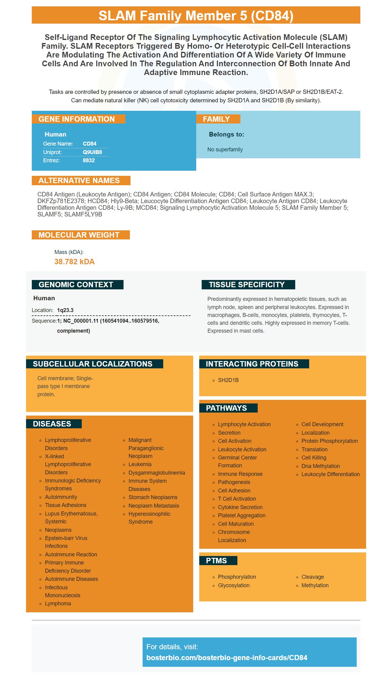
Facts about SLAM family member 5.
Tasks are controlled by presence or absence of small cytoplasmic adapter proteins, SH2D1A/SAP or SH2D1B/EAT-2. Can mediate natural killer (NK) cell cytotoxicity determined by SH2D1A and SH2D1B (By similarity).
Human | |
---|---|
Gene Name: | CD84 |
Uniprot: | Q9UIB8 |
Entrez: | 8832 |
Belongs to: |
---|
No superfamily |
CD84 antigen (leukocyte antigen); CD84 antigen; CD84 molecule; CD84; Cell surface antigen MAX.3; DKFZp781E2378; hCD84; hly9-beta; leucocyte differentiation antigen CD84; leukocyte antigen CD84; Leukocyte differentiation antigen CD84; Ly-9B; mCD84; Signaling lymphocytic activation molecule 5; SLAM family member 5; SLAMF5; SLAMF5LY9B
Mass (kDA):
38.782 kDA
Human | |
---|---|
Location: | 1q23.3 |
Sequence: | 1; NC_000001.11 (160541094..160579516, complement) |
Predominantly expressed in hematopoietic tissues, such as lymph node, spleen and peripheral leukocytes. Expressed in macrophages, B-cells, monocytes, platelets, thymocytes, T-cells and dendritic cells. Highly expressed in memory T-cells. Expressed in mast cells.
Cell membrane; Single-pass type I membrane protein.
Are you aware of the best uses for the CD84 marker? This article will provide information on how CD84 is expressed during fibrosis and how you can benefit from it. In addition, you'll learn how this marker is used to diagnose and treat the condition. Here are some tips for using the CD84 marker:
You should test your blood for markers like Boster Bio CD84, if you think you might be suffering from fibrosis. There are a variety of tests that can detect this fibrosis marker. Some markers are secreted while others are transmembrane and extracellular. The tests also check for BMI albumin, BMI, as well as ALT levels. These are all markers that are linked to fibrosis.
These markers are commonly used to diagnose a range of diseases and disorders. They are useful in diagnosing liver fibrosis, alcohol-induced liver steatohepatitis as well rejection of transplants. They are also useful in monitoring the progress of disease. They can also be used to detect liver disease and NASH. They can also be utilized to monitor the progress of disease and to track fibrosis of other organs.
It is crucial to detect the fibrosis-related markers for treatment. There are a variety of methods used to identify this marker, including mass spectrometry, NMR, microfluidics, immunoassays, ELISAs, and aptamers. These methods can detect the presence of a specific marker in blood and also monitor the disease's progression. This information can be used to inform treatment decisions since the level of the marker can indicate the likelihood of further disease progression.
In addition to CD84 Boster Bio's fibrosis panels contains 61 proteins. The most well-known protein in this panel is CD84. It helps identify the presence of markers in the blood of a particular patient. The results can be used to assist doctors determine the best treatment for their patients. For instance, if Boster Bio CD84 is found in blood samples, it could be an indicator of fibrosis in patients with liver fibrosis.
The presence of inflammatory macrophages can be detected by the CD84 marker. This condition is characterized by an increase in fibroblasts as well as myofibroblasts. Fibroblasts are inflammatory cells that serve a variety of functions. They are responsible for recruiting neutrophils to secrete protein and promoting cell growth. This marker has been discovered to be highly expressed in COVID-19 lung tissue.
Researchers have identified a novel macrophage subpopulation that is present in patients suffering from advanced pulmonary fibrosis. These cells have the CD84 marker more frequently. The results support the notion that this subpopulation is an appropriate target for disease therapy. The new biomarker may be beneficial in the treatment of fibrosis. There aren't any treatments for this subpopulation however, the discovery of this marker could provide a fresh way to treat.
Both diseases exhibit similar changes in the alveolar epithelial cell numbers. The airway epithelial cell is gradually reduced in number despite the fact that alveolar cells are increasing. The reduced number of these cells may be due to increased cell death pathways and a stunted compensatory response. Patients suffering from pulmonary Fibrosis might also have abnormal basaloid cells. These cells are not understood from where they came from and have altered gene expression profiles.
Myofibroblasts are known to be rich in fibrillar as well as other collagen. However their source isn't identified. However, Habermann et al. identified four distinct phenotypes for lung fibroblasts: HAS1hi fibroblasts are found in the subepithelial areas within the airways, the HAS1hi fibrocytes were found in the immediate subpleural region and the PLIN2 and other LUM+ fibroblasts can be found extensively throughout the parenchymal areas. These phenotypes of lung fibroblasts will likely to change in vivo since the population is highly plastic.
Research has also proven that IL-21A and IL-17A could play a significant role in fibrosis of the liver. The Smad signaling pathway may be an immune therapeutic therapy for treating liver fibrosis. While further research is required to determine if these proteins are involved in liver fibrosis development, the study offers a promising start.
In mice, CD4+ Tfh cells exhibit distinct gene signatures which are independent of Th27 and Th2 cell lines. CD4+ Tfh cells are involved in the inflammation and fibrosis of the liver caused by schistosome-induced disease. These immune cells also have high levels of ICOS which is linked to inflammatory conditions. Liver fibrosis is also affected by IL-21 and PD-1 levels.
The Boster Bio CD84 marker is a novel protein that is expressed by a variety of cells and is used in the diagnosis, treatment, and screening for fibrosis. This antibody is used to determine cells that express CD84Hy1 in biological samples. This antibody is effective in identifying diseases like cancer, fibrosis, organ transplant rejection and allergic reactions.
This protein is found in several autoimmune disorders that include rheumatoid and Lupus erythematosus. It is also found in the liver, kidneys and in the hearts that are rejected. For patients suffering from autoimmune disorders it suppresses the immune system by targeting the cells that express it. The cells then begin to die. To prevent fibrosisfrom occurring, it is crucial to identify the onset of fibrosis in the early stages.
The Boster Bio CD84 marker is also able to be conjugated to radioligands and toxins. By targeting the CD84Hy1 protein, a treatment agent may be directly delivered to tumor cells to prevent further damage. This antibody may also be utilized as an instrument for diagnosing. The anti-CD84Hy1 antibody can be used to determine if fibrosis is present. It is safe to use during radiotherapy, chemotherapy, and surgery.
The progression of hepatic fibrosis is affected by IL-21+ cells located in the liver. They are found in the liver disse gap and express GFAP and IL-21. An increase in IL-21R levels may lead to liver fibrosis. The protein is found on both immune and nonimmune cells. It is also a possible target in the treatment of hepatic Fibrosis.
Boster Bio CD84 comes from an herpes simplex virus or influenza virus. These peptides are used to immunize animals against these antigens. The antigens can then be delivered to T cells within antigen presenting cells. This triggers an immune reaction against CD84Hy1. U.S. Pat. describes the methods used to synthesize. No. 6,300,090.
PMID: 9310491 by de la Fuente M.A., et al. CD84 leukocyte antigen is a new member of the Ig superfamily.
PMID: 10698700 by Krause S.W., et al. Characterization of MAX.3 antigen, a glycoprotein expressed on mature macrophages, dendritic cells and blood platelets: identity with CD84.