This website uses cookies to ensure you get the best experience on our website.
- Table of Contents
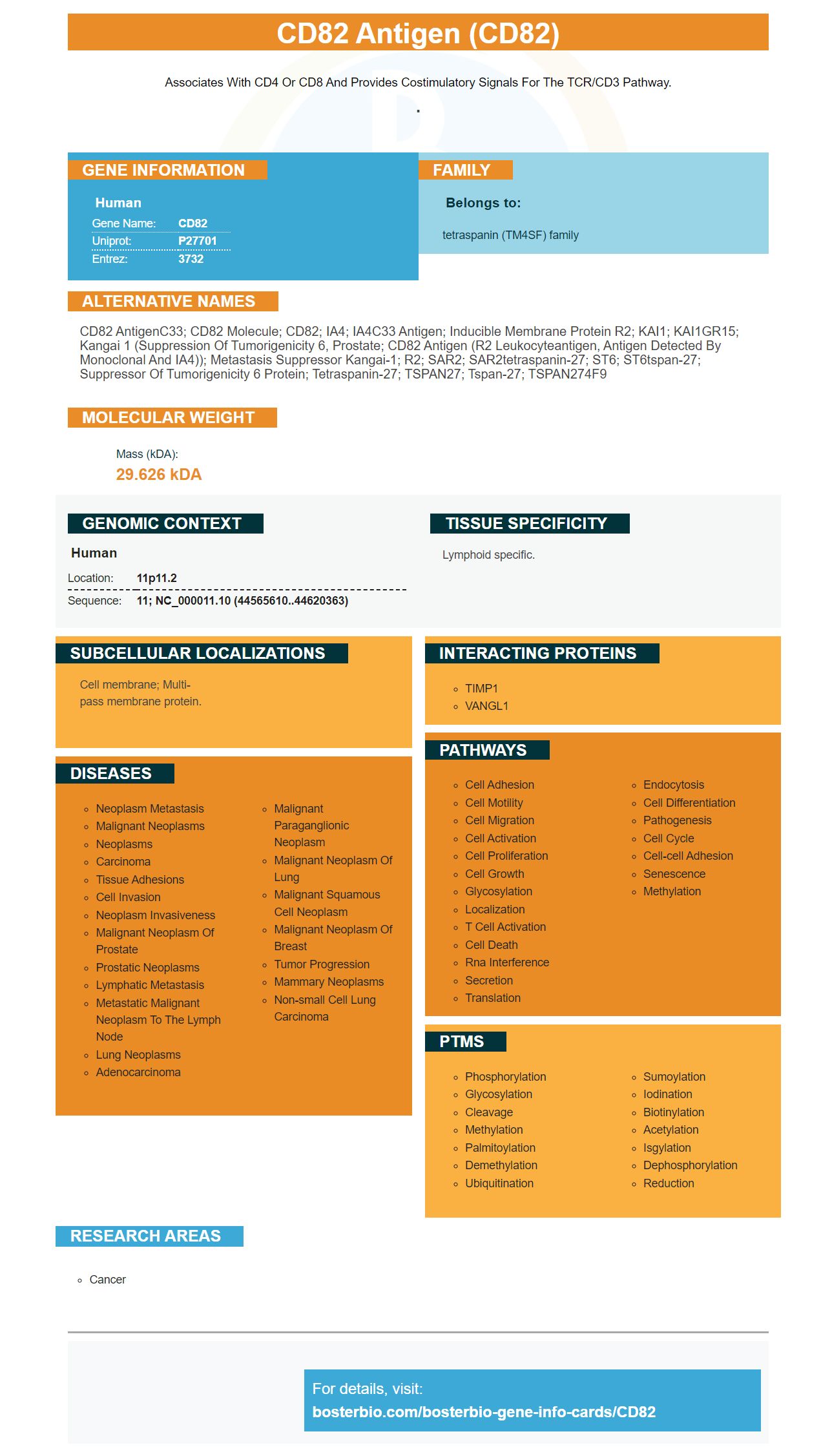
Facts about CD82 antigen.
Associates with CD4 or CD8 and Provides costimulatory signals for the TCR/CD3 pathway.
.Human | |
---|---|
Gene Name: | CD82 |
Uniprot: | P27701 |
Entrez: | 3732 |
Belongs to: |
---|
tetraspanin (TM4SF) family |
CD82 antigenC33; CD82 molecule; CD82; IA4; IA4C33 antigen; Inducible membrane protein R2; KAI1; KAI1GR15; kangai 1 (suppression of tumorigenicity 6, prostate; CD82 antigen (R2 leukocyteantigen, antigen detected by monoclonal and IA4)); Metastasis suppressor Kangai-1; R2; SAR2; SAR2tetraspanin-27; ST6; ST6tspan-27; Suppressor of tumorigenicity 6 protein; Tetraspanin-27; TSPAN27; Tspan-27; TSPAN274F9
Mass (kDA):
29.626 kDA
Human | |
---|---|
Location: | 11p11.2 |
Sequence: | 11; NC_000011.10 (44565610..44620363) |
Lymphoid specific.
Cell membrane; Multi-pass membrane protein.
When it comes to choosing a high-quality primary antibody, Boster is an excellent choice. Their antibodies are validated on multiple platforms and have been extensively tested on Western Blotting, Immunohistochemistry, and ELISA. Boster also rewards its first reviewers with product credits and the chance to win one of the products. They also reward scientists world-wide for their contributions to the scientific community.
The primary antibodies developed by Boster have been cited in research literature for over 25 years. These antibodies have been validated in immunohistochemistry, Western Blotting, and ELISA. This high citation rate is a testament to the quality of the antibodies, which are widely used in clinical trials. However, citation rates alone do not determine the quality of primary antibodies. To determine if a primary antibody is cited in a research paper, it must be based on several different factors, including the research project itself, as well as the subject of the article.
The primary antibodies produced by Boster are remarkably effective. These include the IgA, Type I collagen, HTATIP, and Monocyte/Granulocyte PE, which are largely used in cancer research. The BRCA1 and HTATIP antibodies are particularly useful in identifying breast cancer, as they target the BRCA gene. In addition, these antibodies also target PARP10 and IDh2, which are members of the poly (ADP-ribose) polymerase family. These antibodies also target the anti-viral virus VSV-M.
To ensure the highest degree of sensitivity and specificity, Boster Bio CD82 markers undergo thorough validation on Western Blotting, Immunohistochemical, and ELISA methods. These techniques use primary antibodies that bind to a target protein, followed by labelled secondary antibodies. These methods allow the detection of very small quantities of protein and assess its molecular weight.
Each Boster Bio CD82 marker has been rigorously validated on Western Blotting, Immuno-histochemistry, and ELISA. Each of these three methodologies consists of several steps, including sample preparation, antibody concentration, and blocking buffer. Boster Bio optimization guides and tips answer all your questions regarding ELISA and Western Blotting and help you optimize your experiments.
This article was written under the Creative Commons Attribution License (CC-BY-SA). It can be used in other publications or forums as long as it is cited and the original authors are credited. The authors declare no financial or commercial relationships with any of the companies mentioned. The data presented in this article were obtained from multiple studies, which accounted for a diverse range of samples.
In addition to Boster Bio CD82 markers, these immunoassays have been extensively validated on Western Blotting, Immuno-histochemistry, and ELISA. In addition, the Boster Bio CD83, CD33, and CD90 were shown to be inversely correlated with Gleason scores of HB and were correlated with various clinicopathological parameters. The expression of these markers is associated with a decreased overall survival and a reduced DFS compared to early and advanced adenocarcinoma.
Additionally, Boster Bio CD82 markers are a high quality product. Each marker has been thoroughly validated on Western Blotting, Immunohistochemistry, and ELISA. Boster Bio CD82 markers are highly sensitive, specific, and highly accurate. Their reliability and accuracy have been demonstrated in studies involving human tumor samples and cell lines.
In a study to evaluate the efficacy of Boster Bio CD82 as a Western Blotting marker, researchers used a lentivirus expressing a V5-tagged version of CD82. The lentivirus was used to infect normal primary cells and DMD myoblasts. The resulting cultures were analyzed for EdU incorporation. There were no significant differences between CD82 overexpressing cultures and controls. Moreover, DMD myoblasts intrinsically incorporated a greater amount of EdU than normal cells. And, the expression of MRF4 was significantly higher in CD82-overexpressing cells compared to controls.
To ensure a beautiful and clear Western blot, sample preparation is critical. Protein extraction methods vary in effectiveness and can be the difference between a blank blot and a stunningly beautiful one. Boster provides specialized extraction kits as well as lysis buffers to suit any need. Fortunately, Boster has a comprehensive guide for choosing the right extraction method for your experiment. Transfer methods differ in the amount of equipment needed, the speed and the efficiency of the process. Therefore, it is vital to choose the correct method.
The Boster Bio CD82 series of myogenic cell antibodies is designed to detect expression of this protein in the cell membrane of mononuclear myogenic cells. Myogenic cells express MyoD and dystrophin and express CD82 as a cell-surface protein. Using these antibodies, scientists can monitor the progression of DMD by studying the protein's function. For further studies, this antibody is highly suitable for immunofluorescence experiments.
The Boster CD82 antibody is available in a variety of formats, including a variety of cell types and assays. Its high-affinity antibodies have been widely cited in the research community for over 25 years, and are suitable for immunofluorescence, immunohistochemistry, and ELISA applications. It is available in a range of species, including rabbit polyclonal antibodies and mouse monoclonal antibodies. In addition, Boster offers free secondary antibodies with every purchase.
A number of functional studies have shown that Boster Bio CD82 markers are highly effective for immunofluorescence. In addition, the CD82-shRNA mice were found to have significantly decreased myogenic activity, while their counterparts expressed more MRF4. Furthermore, a significant decrease in the proliferation of CD82-shRNA mouse myoblasts was found in DMD myoblasts, whereas there was no change in the proliferation of normal muscle cells.
The Boster Bio CD82 antibodies were developed with high precision for use in immunofluorescence experiments. Their compatibility with immunofluorescence is confirmed by the PLA assay. The Boster Bio CD82 antibodies are designed for use in immunofluorescence experiments, and they can be used to detect the a-sarcoglycan protein complex in muscle cells.
CD82 is a member of the tetraspanin family. They play an important role in metastases. They can either suppress or promote metastasis. Transfection of tetraspanins in cancer cell lines reduced their metastatic potential. The reduced expression of these proteins correlates with a reduced survival rate, as shown in several clinical studies. When CD9 and CD82 protein levels are reduced, metastases are more likely to occur.
IHC is essential in many research fields. By labeling specific components of cells and tissues, researchers can identify and track their functions. Several methods are used to achieve this. Direct immunofluorescence staining uses antibodies that are directly conjugated to the fluorescent tag. The secondary antibody may fail to penetrate the cells because of the large antibody-fluorophore complexes. Indirect immunofluorescence requires the use of secondary antibodies.
PMID: 1842498 by Gaugitsch H.W., et al. A new superfamily of lymphoid and melanoma cell proteins with extensive homology to Schistosoma mansoni antigen Sm23.
PMID: 1401919 by Imai T., et al. C33 antigen recognized by monoclonal antibodies inhibitory to human T cell leukemia virus type 1-induced syncytium formation is a member of a new family of transmembrane proteins including CD9, CD37, CD53, and CD63.