This website uses cookies to ensure you get the best experience on our website.
- Table of Contents

92 Citations 18 Q&As
45 Citations 4 Q&As


25 Citations 16 Q&As


Facts about Hematopoietic progenitor cell antigen CD34.

Presents carbohydrate ligands to selectins. .
| Human | |
|---|---|
| Gene Name: | CD34 |
| Uniprot: | P28906 |
| Entrez: | 947 |

| Belongs to: |
|---|
| CD34 family |

CD34 antigenhematopoietic progenitor cell antigen CD34; CD34 molecule; CD34; HPCA1
Mass (kDA):
40.716 kDA

| Human | |
|---|---|
| Location: | 1q32.2 |
| Sequence: | 1; NC_000001.11 (207880972..207911125, complement) |
Selectively expressed on hematopoietic progenitor cells and the small vessel endothelium of a variety of tissues.
Membrane; Single-pass type I membrane protein.


The best method of finding the best Anti-CD34 antibody for your research is to search for a specific one that will target this marker. These antibodies may not be effective because they are not specifically enough or have significant variations from batch batch. This is particularly relevant for immunoblotting and CD34 sorting. It is recommended to stick to monoclonal antibodies to accomplish these tasks.
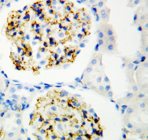
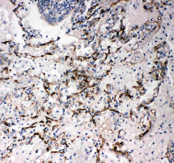
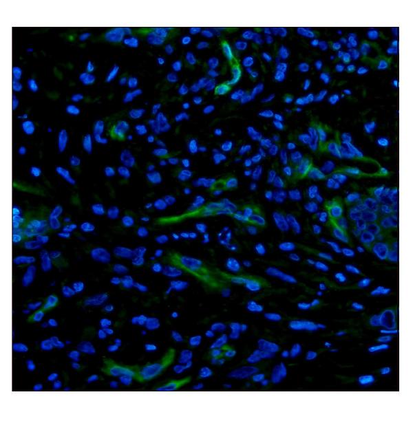
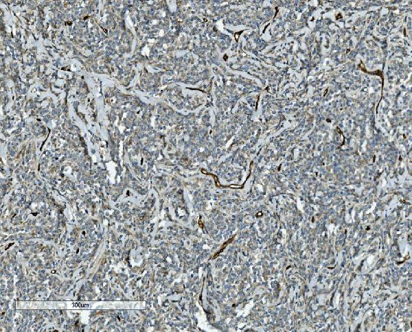
Anti-CD34 antibody from Boster Bio is a rabbit monoclonal antibody which recognizes CD34. CD34 is a monophosphoprotein of type I with a molecular weight of between 105 and 120kD. Humans rodents, mice, and humans all express this protein, and it is useful in immunocytometry, immunocytochemistry and immunofluorescence. The bosterbio antibody was developed with the use of mice and rabbits for research.
This highly specific antibody has been validated in IHC, Flow Cytometry, and Western Blotting. Boster also offers ELISA kits that detect biomarkers in immunohistochemistry, development biology, and neurosciences. Boster's antibodies are validated using untransfected cell lines to ensure precise results. Boster Bio also offers technical support for its antibodies. They are available to purchase through Tebu-bio.
The Antibody Resource can help you determine whether or whether to purchase the Boster Bio Anti-CD34 anti-CD34 antibody. They also offer Superstarter kits that come with trial-size versions of their top-rated CD34 antibodies. These small-sized antibodies can be used in conjunction with certain experimental conditions to determine the best antibody for your research. You can also purchase a pre-made kit to determine whether the antibody you're interested in reacts with the target.
In vitro studies have demonstrated that keratocytes display characteristics of MSC. However the absence of CD34 suggests that they have resigned themselves to a lineage. A large proportion of CD34+ cells express tissue-specific markers such as epithelial progenitors and keratocytes live. However, this does NOT mean that keratocytes do not have MSC.
Although the expression of CD34 has been recognized as a common marker for progenitors, it has not been studied across all cell types. Different studies have demonstrated that CD34 is expressed on several kinds of cells, such as embryonic fibroblasts and endothelial cell. Understanding the connections between different types of cells will help us understand the role of CD34. It has been established that CD34+ cells are more able of differentiation than cells without the marker.
Although CD34 expression is more prevalent in muscle cells than it is on myogenic precursors of all kinds however, it has not become more prevalent. The expression was first detected after the emergence of committed muscle satellite cells, indicating that CD34+ satellite cells were already committed to a myogenic fate. CD34satellite cells that are + are more likely than those without this marker to be myogenic. If this is the scenario, CD34 expression can be an effective marker for myogenic differentiation.
While the expression of CD34 is seen throughout the body, the majority of it has been discovered in skin tissue. CD34 is a crucial component in keratocytes and may help to identify a proliferating set of microglia within spinal cords. Additionally, the expression of CD34 in skin tissue is usually seen in areas that are damaged by diseases such as stroke and cancer.
The uses of the CD34 marker are many and diverse. It is used in medicine to restore hematopoiesis after the treatment of marrow. Traditional colony-forming tests take between ten and fourteen days to complete and are therefore not suitable for immediate evaluation of HPC products. It is also difficult to determine CD34+ HPCs, as it is often obscured or deformed by non-specific populations. This resulted in a wide variation in the recommended CD34 values for long-term transplantation.
Despite its widespread use as a progenitor cell marker, CD34 is not well studied in all cells. In actuality the marker is expressed by a variety of types of cells that include embryonic fibroblasts, multipotent mesenchymal stromal cell types epithelial progenitors and a variety of other types of stem and progenitor cells. The applications of this marker are restricted, however, outside of the specialty areas of medicine. Its expression is also limited to these cells, which is the reason these cells are often considered hematopoietic contaminants.
As far as cancer is concerned, CD34 has been implicated in a myriad of cellular forms that include preB-ALL, AML M7 and alveolar soft Sarcoma. The protein is also present in liposarcoma, gastrointestinal tumors and the granulocytic Sarcoma. In the end, the CD34 marker is a useful biomarker for the hepatic fibrosis.
Future research will allow make use of hES-CD34 cells that come from specific individuals. In addition to HIV studies, CD34 cells may also be used to produce other differentiated cells like T and dendritic cells. There is a lot of potential for replacement of cells in a variety of settings. This marker is an important supplement to gene therapy. The availability of this marker and its widespread use in the clinic makes it an important instrument for future medical research.
The CD34 mAb can be used to sort CD34. The monoclonal antibodies are highly sensitive and selective to specific epitopes found on CD34. There are more than thirty CD34 monoclonal antibodies, with each focused on different epitopes within the CD34 antigen. Table 2 lists the different antibodies that were used in various studies. Here, we look at the best uses of CD34 mAbs.
There are numerous functions that CD34 plays in vasculature. It is thought to act as an underlying structure for glycans that are specific to lineages, which enable stem cells to connect to selectins. The activated proteinkinase has been found to phosphorylate the intracellular chain of CD34 antigen. These glycans permit stem cells to bind selectins.
The CD34 gene is located on the surface of progenitor cell progenitors, but does not associate with hematopoietic or endothelial cells. It is extremely rare in peripheral blood , and can be used to determine different progenitor cell populations of various subpopulations. CD34 can be used to identify stem cells as well as progenitor cell subpopulations that can transplant cells into a patient's bodies.
CD34 mRNAs were synthesized with extracts from rat lung. Primers made from the NCBI database's predicted rat CD34 sequence were employed. The PCR product was comprised of two distinct partial sequences 1,161 nucleotides in variant 1 and 1.312 nucleotides in variant 2. These clones were deposited in GenBank.
To develop a monoclonal antibody against CD34, the scientists first determined the expression pattern of CD34 in a variety of human and mouse tissues. They discovered that the full CD34 is expressed in the plasma membranes of luminal endothelial cells in the heart and lung. However, reduced CD34 was more present in kidneys and the liver. Clones are the best use of the CD34 marker
Although the role of the antigen remains being debated however, it is possible to keep the CD34 marker in a simple method to preserve its effectiveness. Cells that have been enriched with CD34 antigen can be stored at 4 degrees C or room temperature in the absence of stirring. It can also be employed as an agent of binding for selectins. A phosphorylation enzyme called activated protein kinase C (Akt) phosphorylates the intracellular chain of the CD34 antigen.
It is not known what the optimal storage conditions for CD34-positive cord blood cells. In the current study, CD34-positive cells were isolated as soon as they were collected and then stored at different temperatures for up to 24 hours, 48 hours, and 72 hours. The CD34 immunophenotype could be evaluated by flow cytometry. CD34 positivity remained unchanged after storage for 24 hours, 48 hours, and 72 hours. CD45+ positivity, however, decreased regardless of storage temperature.
A biochemical test for GUSB activity showed that it was widely distributed throughout the tissues of NOD/SCID/MPSVII animals at 12 and 6 weeks. This suggests that the bone marrow remained a reservoir of storage material after transplantation. The researchers also found that CD34-positive cells are present in the bone marrow where they can be stored in a way that is functional. If the storage material within the bone marrow of your body is not properly stored, it could worsen your health.
The NOD/SCID/MPSVII mouse modeling was created to assess the in vivo locations and effects of treatments in a realistic model of the disease. The mice could be transplanted with human CD34+ precursor cells of hematopoietic origin. This allows them to assess the effectiveness of therapeutic strategies against Lysosomal Storage Disorder. The mice used in these studies were able produce CD34+ cells and the resulting effect was similar to the effects observed in NOD/SCID/MPSVII mice.
PMID: 1370171 by Simmons D.L., et al. Molecular cloning of a cDNA encoding CD34, a sialomucin of human hematopoietic stem cells.
PMID: 1374051 by Satterthwaite A.B., et al. Structure of the gene encoding CD34, a human hematopoietic stem cell antigen.
*Showing only the more recent 20. More publications can be found for each product on its corresponding product page