This website uses cookies to ensure you get the best experience on our website.
- Table of Contents
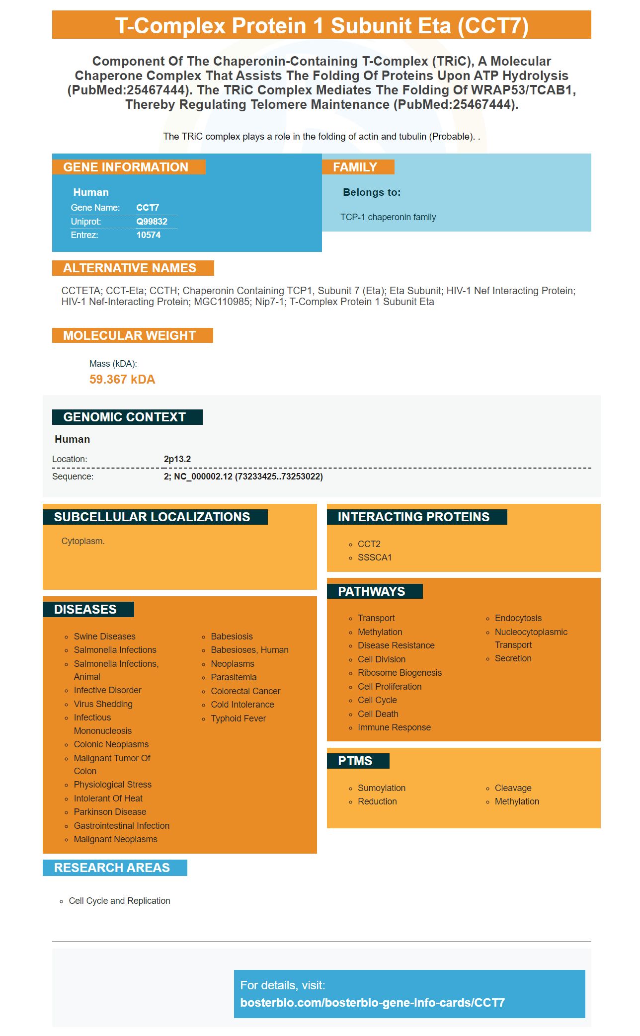
Facts about T-complex protein 1 subunit eta.
The TRiC complex plays a role in the folding of actin and tubulin (Probable). .
Human | |
---|---|
Gene Name: | CCT7 |
Uniprot: | Q99832 |
Entrez: | 10574 |
Belongs to: |
---|
TCP-1 chaperonin family |
CCTETA; CCT-eta; CCTH; chaperonin containing TCP1, subunit 7 (eta); eta subunit; HIV-1 Nef interacting protein; HIV-1 Nef-interacting protein; MGC110985; Nip7-1; T-complex protein 1 subunit eta
Mass (kDA):
59.367 kDA
Human | |
---|---|
Location: | 2p13.2 |
Sequence: | 2; NC_000002.12 (73233425..73253022) |
Cytoplasm.
This article will discuss the autoradiography film, the DAB chromogenic detection system, the protein transfer efficiency using membrane staining and the ECL chemiluminescent detection system. Steven Boster discovered CCT7 in 1993. This marker is now widely used in many medical fields. Boster Bio: Best Uses Of The CCT7 Marker
The boster Bio.CCT7 marker is an antibody that has a high affinity against CCT7. It has been validated for ELISA, immunohistochemistry, and Western blotting. Boster also provides high-affinity prima antibodies. These antibodies are trusted and respected by the research community. You can find a complete list of Boster Bio antibodies. Most of their products come in a variety concentrations.
The ECL chemistry is a fast and effective way to analyze protein expression in biological samples. It can detect the immunoreactivity of a protein to identify highly expressed proteins. ECL uses a small amount (or none) of protein to detect antigens. The system can be used with many types of samples, including cell membranes and tissue sections as well as serum. Boster Bio and several other vendors sell the ECL kits.
The ECL method uses antibodies conjugated to HP to detect proteins. At 450nm, strong blue emission occurs as the enzyme reacts on the substrate. Unlike other detection methods, light emission occurs only during the enzyme-substrate reaction, and the signal output ceases as soon as the substrate is exhausted. Before performing an analysis, it is important to examine the conditions of each reaction.
Use 10 mM Tris pH 7.4 @ 22°C to make the sample buffer. Additionally, you can use 0.5 mM Tris pH 7.4 at 22 degC, 1 mM NAH2PO4, 0.2% SD, and 1% Tween-20. The membrane should be rinsed with Milli-Q or double-distilled water after the wash. Be sure to gently stir the sample during rinsing so that the reagent can be applied.
The optimal enzyme to substrate ratio is what makes western blotting the best. Because of its high sensitivity enhanced chemiluminescence has been a preferred method of detection for many researchers. The system also provides high signal-to-noise ratio and wide dynamic range. ECL technology is widely used in biomedical and scientific research. Its simple and cost-effective design makes it easy integrate into lab workflows.
Clarity Max are compatible to any horseradish oxidase conjugate. This makes them suitable for most western blot applications. They are also good for optimized westernblot experiments, which require a lot of proteins. This ECL substrate must be kept from direct sunlight for prolonged periods of time. Once exposed to sunlight, the signal can be clearly seen on X-ray film and digital images.
The DAB chromogenic detection system is a powerful Intensification kit that integrates DAB and Streptavidin-peroxidase staining methods. This system uses horseradish-peroxidase as a catalytic agent to catalyze the laying of two distinct Linkers. The one that binds with the target is the linker, while the other binds with the conjugate. The DAB chromogenic detect reagents produce a highly sensitive, specific chromogenic sign.
The nature of your membrane, the molecularweight of the protein and the type thereof all influence the efficiency of protein transfer. Higher molecular weight proteins have higher transfer rates to the membrane. Low-abundance genes can be difficult for people to identify. These issues affect the reproducibility and sensitivity to WB results. Boster Bio membrane staining improves protein transfer efficiency.
In two separate studies, the efficiency of protein transfer was also examined. First, a protein mix was separated with 8% SDS/PAGE and transferred onto either PVDF or NC membrane. The samples were then stained by different antibodies. After the membrane was scanned, the staining intensity was calculated. AAL and ApoA1 are the most commonly used antibodies in this test. These two antibodies are used to identify the proteins present in the sample.
The second method for measuring protein transfer efficiency within a cell culture system is electrophoresis. The gel is sandwiched between a PVDF membrane and a nitrocellulose membrane. To prevent air bubbles from forming, these membranes were clamped together. The membrane was then immersed into a buffer and an electricity field. A Coomassie Brilliant Blue R-250 dye was used to compare the levels of protein binding to the membrane to the levels of transfer in the samples.
A membrane compatible with the sample is required for electrophoresis. The membrane used should have pores that vary in size according to the protein molecular weight. Smaller pores will allow the protein to be combined tightly with a membrane of larger size. The PVDF membrane is also more sensitive, responsive, and able to combine proteins with larger membranes than the NC membrane. These factors increase the detection of small-sized molecules within the cell membrane.
Preparing the gel or membrane sandwich is the first part of this procedure. You can remove air bubbles from the gel using a pipette, or a roller. You can also place the sandwich in a transfer buffer dish. Although the membrane should be the same in size as the paper, large overhangs may prevent current from flowing through it. If all three steps have been completed successfully, the sample will be visible in the gel as one protein band.
PMID: 9819444 by Won K.-A., et al. Maturation of human cyclin E requires the function of eukaryotic chaperonin CCT.
PMID: 14532270 by Imai Y., et al. A product of the human gene adjacent to parkin is a component of Lewy bodies and suppresses Pael receptor-induced cell death.