This website uses cookies to ensure you get the best experience on our website.
- Table of Contents
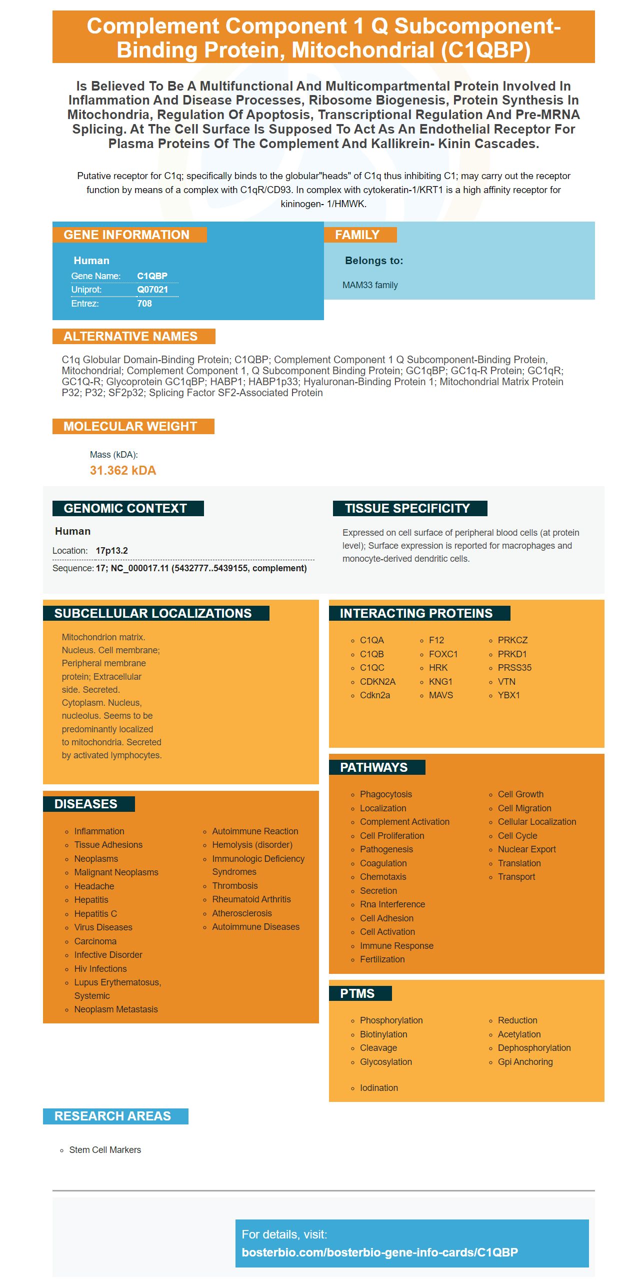
Facts about Complement component 1 Q subcomponent-binding protein, mitochondrial.
Putative receptor for C1q; specifically binds to the globular"heads" of C1q thus inhibiting C1; may carry out the receptor function by means of a complex with C1qR/CD93. In complex with cytokeratin-1/KRT1 is a high affinity receptor for kininogen- 1/HMWK.
Human | |
---|---|
Gene Name: | C1QBP |
Uniprot: | Q07021 |
Entrez: | 708 |
Belongs to: |
---|
MAM33 family |
C1q globular domain-binding protein; C1QBP; complement component 1 Q subcomponent-binding protein, mitochondrial; complement component 1, q subcomponent binding protein; gC1qBP; GC1q-R protein; gC1qR; gC1Q-R; Glycoprotein gC1qBP; HABP1; HABP1p33; Hyaluronan-binding protein 1; Mitochondrial matrix protein p32; p32; SF2p32; splicing factor SF2-associated protein
Mass (kDA):
31.362 kDA
Human | |
---|---|
Location: | 17p13.2 |
Sequence: | 17; NC_000017.11 (5432777..5439155, complement) |
Expressed on cell surface of peripheral blood cells (at protein level); Surface expression is reported for macrophages and monocyte-derived dendritic cells.
Mitochondrion matrix. Nucleus. Cell membrane; Peripheral membrane protein; Extracellular side. Secreted. Cytoplasm. Nucleus, nucleolus. Seems to be predominantly localized to mitochondria. Secreted by activated lymphocytes.
If you are looking for an antigen that is primary that you can use, you should look into Boster Bio. Boster Bio is well-known for its high-affinity primary antibody. Boster Bio also offers Anti-Complement 4d which can be used to conduct studies in a variety of cancerous tissues. You can find out more about this antibody by reading our article.
Flow Cytometry, which employs proteins to analyze biological samples is a significant method. Samples can be cells or particles. Boster Bio provides high-affinity monoclonal as well as polyclonal primary antibody. Boster Bio's antibodies have received numerous reviews and will continue to receive them. Here's a summary of the products offered by Boster. Find out more about Boster's unique capabilities.
Established in 1993, Boster Bio is a company which specializes in high-affinity sensitivity, and specific antibodies. Over the past two decades of study and development have gone into the company's process of enhancing its technology. Boster Bio's antibodies are used in more than 29,000 scientific publications and are fully verified for WB, ELISA and Flow Cytometry. A majority of their products have been subjected to rigorous screening against a variety of samples to determine their quality.
When determining the specificity of an antibody in determining its specificity, the International Working Group for Antibody Validation recommends that it be validated using multiple cell types and tissues. High-affinity antibodies are produced by genetic strategies that ensure the antibodies don't give an indication in knockout mice of the target protein. The specificity of an antibody can be tested by creating antibodies against multiple epitopes of the same protein.
Another company that makes monoclonal and polyclonal antibody is Abcore. Abcore is USDA and NIH-certified and uses rabbits and chickens to produce. They provide custom peptide designs, production, and purification of mouse monoclonals. Their team is equipped to tackle the most complex antibody projects, including those that require large quantities of monoclonal antibodies.
An antibody that detects this protein is called the acute the hematologic rejection marker C4d. The C4A antigen was used to construct the antibody. This monoclonal antibody reacts with Human C4d. It has been proven effective for Immunofluorescence. Boster Bio Anti-Complement 4d Acute Humoral Rejection Marker is available in a 10 mM PHBS with 0.05 percent BSA. It has been tested for use in WB or ELISA.
C4d is a surface-bound protein that is not functional as a C3 convertase. It is broken down by factor I within the bloodstream. C4d that is bound to the surface acts as an AMR marker. C4d is not a specific protein, however it does not possess any biological functions or receptors. It binds with tissue close to the location where complement activation occurs.
C3 deficiencies can hinder the immune response to allogeneic transplants. The mice with C3 deficiency have a longer survival time of skin grafts. The evidence from clinical studies also indicates that C4d deposition within the capillaries of the peritubular can be associated with poor clinical outcomes. This finding is consistent with other studies.
The Ab is extracted from the Bioreactor Concentrate with Protein A/G. It is made in 10mM PBS containing 0.05% BSA or azide. Its concentration is 1 mg/ml. It is a non-diagnostic Ab is not approved for diagnostic use. It is essential to speak with a doctor if you require an exact test.
C3d is a protein that has an approximate molecular mass of 210 kg. It can be found in human serum at concentrations up to 600 mg/ml. It consists of three subunits with a molecular weight of 80 kDa, 90 kDa and 30 kDa. They are connected by disulfide bonds. C4d is a product split of C4 and is created in the process of activating complement. C3d's biological activity is controlled by an feedback loop that is positive, which causes local B-cell proliferation and production of antibodies.
The Acute Humoral Rejection Marker can be used to diagnose and treat patients. C3d levels must be checked for elevated C4d levels in patients suffering from acute renal rejection. Furthermore C4d antibody C4d antibody can help the physician determine the cause of rejection of allografts.
One of the most important functions of C1q is to regulate the epithelial-mesenchymal transition (EMT). It binds with a variety of cell surface receptors, including C1qRp, alpha2 beta-1 Integrin, and LAIR-112 Protein. C1q can be bind by a variety of kinds of cells, but it's not yet clear how these proteins trigger cellular responses to it.
To determine if C1q promotes EMT and induce invasiveness in HepG2 cells We used a 6-well transwell plate. For 18 hours HepG2 cells were placed in medium containing or without C1q. Cells were placed in the top chambers using an insert for transwells. The insert was covered with a Matrigel-based membrane. After eight hours, cells that had undergone migration were able move through the Matrigel matrix and stick to the bottom membrane. The bottom of the insert was stained with 0.1 percent crystal violet to count invading cells. These cells were then examined against controls.
Cancer-associated fibroblasts release ten times more inflammatory adipocytokines than normal fibroblasts. These cytokines include RANTES and the IL-6. These proteins are found in sufficient amounts in areas of high metabolism, for instance, tumor tissue. This suggests that C1q enhances EMT and triggers the an invasion or migration of these cells.
C1q plays a role in plasma complement production, including C1q. Inflammatory responses and the spread of cancer cells are likely to be controlled by these complement proteins. They also play a role in many aspects of liver function. C1q is also thought to play an important role in the innate immune system. Additionally, it was identified to interact with the ligand DDR1 which aids in the migration and invasion of HepG2 cells.
The cells were maintained in 12-well plates. After 2 h, they were enriched with C1q. The cells were then placed at 37 degrees Celsius in a humidified 5 percent CO2 incubator. To examine the expression levels of C1q and C1q-related proteins, the respective amounts of proteins were immunoprecipitated . They were analysed using western blot.
The interaction between C1q and DDR1 in HepG2 cells was further studied using co-immunoprecipitation and Western blotting. In both studies, C1q binds DDR1, a transcription factor which is essential for HepG2 cell migration. C1q also activates MAPK signaling in HepG2 cells and pharmacological inhibition particular MAPKs affected the migration and invasion of these cells.
Cell migration and invasion are affected by the interaction of C5aR1 and DDR1 and E–cadherin. Both proteins are involved in EMT and aid cancer cells in escaping their primary locations. C5aR1 and E-cadherin have been found to be negatively correlated with one another in lung primary tumors. C1q and Ecadherin-related interactions are also linked to the decrease in invasion and migration of pancreatic cancer cells.
PMID: 8262387 by Honore B., et al. Cloning and expression of a cDNA covering the complete coding region of the P32 subunit of human pre-mRNA splicing factor SF2.
PMID: 8195709 by Ghebrehiwet B., et al. Isolation, cDNA cloning, and overexpression of a 33-kD cell surface glycoprotein that binds to the globular 'heads' of C1q.