This website uses cookies to ensure you get the best experience on our website.
- Table of Contents
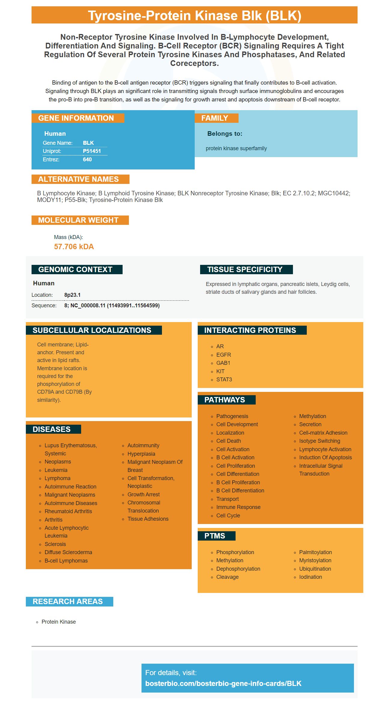
Facts about Tyrosine-protein kinase Blk.
Binding of antigen to the B-cell antigen receptor (BCR) triggers signaling that finally contributes to B-cell activation. Signaling through BLK plays an significant role in transmitting signals through surface immunoglobulins and encourages the pro-B into pre-B transition, as well as the signaling for growth arrest and apoptosis downstream of B-cell receptor.
Human | |
---|---|
Gene Name: | BLK |
Uniprot: | P51451 |
Entrez: | 640 |
Belongs to: |
---|
protein kinase superfamily |
B lymphocyte kinase; B lymphoid tyrosine kinase; BLK nonreceptor tyrosine kinase; Blk; EC 2.7.10.2; MGC10442; MODY11; p55-Blk; tyrosine-protein kinase Blk
Mass (kDA):
57.706 kDA
Human | |
---|---|
Location: | 8p23.1 |
Sequence: | 8; NC_000008.11 (11493991..11564599) |
Expressed in lymphatic organs, pancreatic islets, Leydig cells, striate ducts of salivary glands and hair follicles.
Cell membrane; Lipid-anchor. Present and active in lipid rafts. Membrane location is required for the phosphorylation of CD79A and CD79B (By similarity).
Boster Bio's high affinity primary antibodies have been extensively cited by researchers over the last two decades. These antibodies are used by the scientific community because of their proven performance in ELISA, Immunohistochemistry, and Western Blotting. Continue reading to learn more about Boster Bio's BLK Marker. We would like to hear from you about this topic. So what do you think about Boster Bio's antibodies?
Boster Bio ELISA kits can detect a variety hormonal, small-molecule, oxidative stress, inflammatory cytokines and HSP70. These kits include enough reagents to perform 96 tests. They are also compatible with diverse samples. Boster's kits detect IL-6 and IL-8, IL-10 and EGF.
Flowcytometry has many applications in science, medicine, and the study of cell growth and particle movement. Boster Bio offers a wide variety of high-affinity primary antibodies, both polyclonal and monoclonal, that are highly specific for the applications they are designed for. These antibodies have been used in a variety of applications for over 25 year and are highly cited. They are also available in custom-made kits for use in ELISA applications.
High-affinity primary antibodies from Boster Bio are suitable for use in immunohistochemistry, cell biology, and neuroscience. These kits have been ELISA-validated, and they have been tested against 250 tissue samples. Boster Quality Guarantee is included to back their products. These antibodies are extremely sensitive and specific, so researchers can perform precise tests with confidence.
ELISA testing is used to screen for potential antibodies candidates during antibody development. Following animal immunizations, a titration ELISA is performed on each test-bleed. The hybridoma-fusion method is used to select positive supernatants. The next step is to subclone the wells at the same rate by using limiting-dilution cloning. The selected antibodies are then subjected to further testing.
High-affinity antibodies can be used for enzyme-linked immunosorbent tests (ELISAs). They are sensitive and can detect specific analytes even in crude preparations. However, ELISAs can also produce non-specific results. This is due non-specific binding among primary antibodies. This phenomenon is known as cross-reactivity. It is therefore important to optimize the detection process.
Abnova is just one company that produces antibodies. This company produces monoclonal antibodies in chickens and rabbits, while Abgent focuses on chicken and rabbit polyclonal antibody production. They also specialize on custom antibody development, humanization, Fv fragment optimization, and custom antibody humanization. Biogenes offers contract services for antibody development. All antibodies are tested for mycoplasma contamination, purity, and can be customized for your research.
A nitrocellulose pore membrane is the best choice for protein-blotting. This membrane has a small enough pore to detect low molecularweight proteins. There are many options for nitrocellulose membranes. Which one you choose depends on the type of sample as well as the purpose of the experiment. For more information, please contact a supplier. Here are some of its benefits.
A pooled human serum sample containing 10 mg of protein was resolved using 10% SDS-PAGE. The sample was then electroblotted onto a PVDF or on a nitrocellulose membrane using either a 10mL tube tip box lid or a 10mL pipette. After the membrane has been attached to the membrane, the samples need to be incubated overnight either at 4°C or at room temp. After incubating the samples, it is important to wash the membranes with PBST.
A nitrocellulose membrane is also compatible with western blotting techniques. One hour at 80 mA, proteins are transferred to a Nitrocellulose membrane by using an immobilon PVDF membrane. To detect proteins, a nitrocellulose membrane contains Coomassie Brilliant Blue R-250 dye. A nitrocellulose membrane is highly suitable for Western Blotting.
The nitrocellulose membrane can detect low molecular-weight proteins in a variety bio samples. After fixation, proteins could be detected more efficiently and were retained better. These results prove that the nitrocellulose membrane is a viable option for protein separation. The membrane's sensitivity is up to 16 times higher than PVDF. Further, the membrane is highly resistant to protein binding, which makes it perfect for use in many different applications.
While the binding of proteins to PVDF and NC membranes was similar, CerP's ability to interact with PVDF as well as NC membranes was greater. The protein binding ability to a 15 kD HPB to 0.2 mm NC was greater than that to a 0.45 mm PVDF. These results also indicate which filtration method is best for different applications. If you are looking for a new membrane, you should check out a Boster Bio product today.
Isaac Clarke is one of the many people who have been affected by this BLK Marker. While most people aren’t affected by this strange signal however, some people have been rendered insane or homicidal. Nevertheless, Isaac is largely unaffected by this, suffering only from hallucinations. The BLK marker also creates a dead zone field, which prevents necromorphs coming near the marker. This field keeps necromorphs away from the Marker. It is vital for the Marker because it keeps a steady stream people out on the streets searching for necromorphs.
This resistance makes Isaac a useless asset for the Marker. Isaac is able to hallucinate Nicole because of the Marker. He will have to do everything for Nicole as long as he believes Nicole isn't there. The real Nicole is back at Ishimura. Although the BLK marker is one of the most persistent elements in the Dead Space series, it is not always a threat.
A Necromorph's ability for mutation is a key characteristic of an Infector. It is one of the main causes of death, and the Infectors use the BLK Marker to create a zombie-like body. Infectors can be found near corpses and transform them into Necromorphs in a matter of seconds. Infectors are the only Necromorphs to ignore Isaac.
When the BLK Marker is discovered, Isaac fights Slug with the Asteroid Defence System cannon. The resulting boss fight is one of the most memorable moments in the entire series. The BLK Marker is present throughout the franchise. Isaac must use this to defeat it and save his people. Isaac is the only person who can help a science team investigate the evidence.
PMID: 7822795 by Islam K.B., et al. Molecular cloning, characterization, and chromosomal localization of a human lymphoid tyrosine kinase related to murine Blk.
PMID: 7845672 by Drebin J.A., et al. Molecular cloning and chromosomal localization of the human homologue of a B-lymphocyte specific protein tyrosine kinase (blk).