This website uses cookies to ensure you get the best experience on our website.
- Table of Contents
1 Citations 16 Q&As
8 Citations 17 Q&As


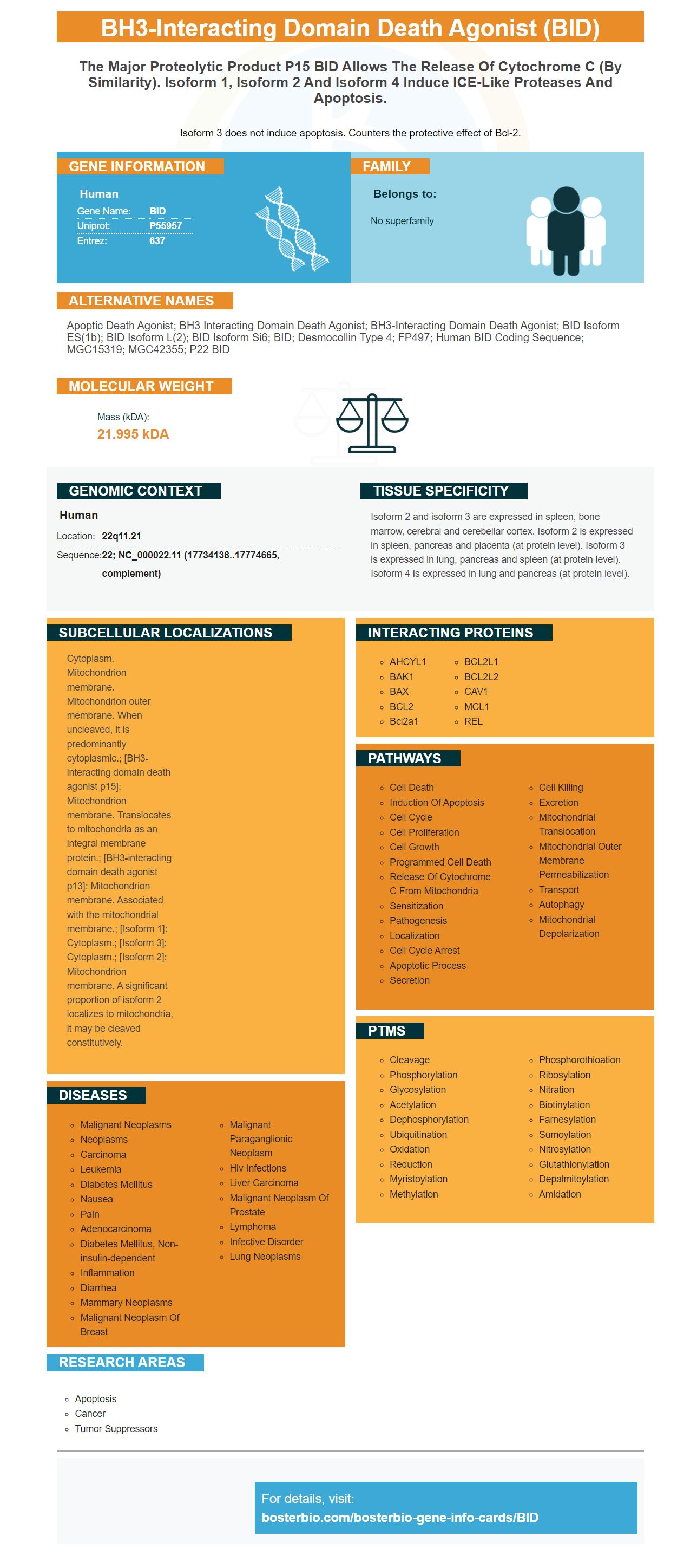
Facts about BH3-interacting domain death agonist.

Isoform 3 does not induce apoptosis. Counters the protective effect of Bcl-2.
| Human | |
|---|---|
| Gene Name: | BID |
| Uniprot: | P55957 |
| Entrez: | 637 |

| Belongs to: |
|---|
| No superfamily |

apoptic death agonist; BH3 interacting domain death agonist; BH3-interacting domain death agonist; BID isoform ES(1b); BID isoform L(2); BID isoform Si6; BID; desmocollin type 4; FP497; Human BID coding sequence; MGC15319; MGC42355; p22 BID
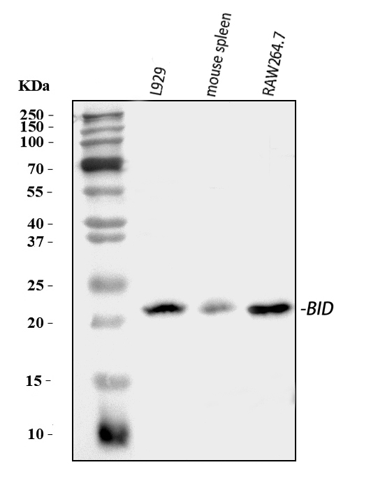
Mass (kDA):
21.995 kDA

| Human | |
|---|---|
| Location: | 22q11.21 |
| Sequence: | 22; NC_000022.11 (17734138..17774665, complement) |
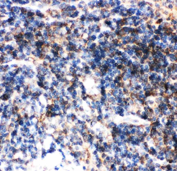
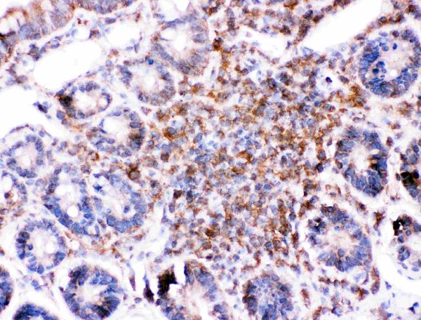
Isoform 2 and isoform 3 are expressed in spleen, bone marrow, cerebral and cerebellar cortex. Isoform 2 is expressed in spleen, pancreas and placenta (at protein level). Isoform 3 is expressed in lung, pancreas and spleen (at protein level). Isoform 4 is expressed in lung and pancreas (at protein level).
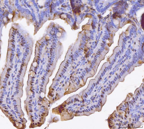
Cytoplasm. Mitochondrion membrane. Mitochondrion outer membrane. When uncleaved, it is predominantly cytoplasmic.; [BH3-interacting domain death agonist p15]: Mitochondrion membrane. Translocates to mitochondria as an integral membrane protein.; [BH3-interacting domain death agonist p13]: Mitochondrion membrane. Associated with the mitochondrial membrane.; [Isoform 1]: Cytoplasm.; [Isoform 3]: Cytoplasm.; [Isoform 2]: Mitochondrion membrane. A significant proportion of isoform 2 localizes to mitochondria, it may be cleaved constitutively.


If you're considering purchasing a Boster Bio, there are numerous things to think about. It offers a clear and illustrated procedure as well as a step-by step IHC protocol. In addition, the guide also contains information on the background and history of the Boster Bio, as well as suggestions and tricks to maximize your research. Whatever your budget you'll find a comprehensive guide to Boster Bio optimization helpful.
An IHC workflow is comprised of the necessary steps to perform a specific immunohistochemistry experiment. Even though the process doesn't require the extraction of proteins from samples, it is essential to prepare the samples for optimal results. The preparation of samples includes fixing, slicing, mounting, and drying. Boster Bio offers a step-by-step IHC protocol that gives guidelines for preparation of the sample and describes the IHC workflow. This step-by step guide contains detailed information about preparation of samples which includes the necessity to thaw frozen samples for greater viability.
The Boster Bio IHC workflow demonstrates the proper use of DAPI that is a commercially-available product from Boster Bio. DAPI was purchased from Wuhan Boster Bio-Engineering Limited Company. The Raji and Jeko cell lines were obtained from Professor Tadashi Yoshino and Professor Tadashi Yoshino. Beijing Weitong Lihua Experiment Animal Technology Co. Ltd. purchased 5 week-old BALB/c female mice.
The Boster Bio A detailed stepwise IHC workflow is provided with examples of each step and discusses the importance of quality control. The manual that goes along with the qScript cDNA synthesizer kit. It also provides guidelines for the process of purifying RNA and sequencing. Boster Bio IHC workflows can be used to carry out more complex workflows.
Boster Bio has provided a thorough step-by-step IHC workflow. It has illustrations of all the steps involved in creating IHC images. BosterBio's IHC workflow has detailed protocols for reagents recommended and troubleshooting guidelines. Boster Bio offers a wide selection of reagents that can meet your research requirements. Boster Bio IHC workflows are a great choice for both novice and experienced researchers.
Boster Bio: A comprehensive step-by-step IH protocol that includes a clearly illustrated IHC workflow written by Dr. David Boster, MD PhD, is a must-have guide for pathologists and immunologists. This guide provides the steps needed to prepare an IHC stain. This Boster Bio resource also gives CEUs for those who have completed the educational materials.
Dr. Nadeau is the recipient of numerous patents. She has also been on several editorial boards for journals with high impact. She is an active member of both the American Thoracic Society and Data and Safety Monitoring Board of the National Heart, Lung, and Blood Institute. Her research in the field of food allergy has been widely acknowledged and acknowledged. She has also contributed to a variety of books. One of her most favored books is A Detail Stepwise IHC Protocol with a Clearly illustrated IHC Workflow
Numerous expected cell populations were identified by single-cell transcriptomic profiling. There were six main clusters including SP-B+ cells, AT1+AQP5+, and ciliated cells. In clusters 0, 2 and 3 of the protocol, there are also basal progenitors (P63+) cells as well as neuroendocrine cells.
Steve Boster's family and friends came first in his life. He was proud of his family and loved them more than everything other things. Steve would be the first to call if your car breaks down at 2 a.m. Steve was always there even in the coldest weather. His generosity was extended to his friends as well. He treated them like family and did whatever could to help. You'll be able to appreciate the importance of family if you've ever in his shoes.
If you are unsure what you can do to improve your immunohistochemistry research, Boster Bio has an easy-to-use guide to answer your questions. No matter if your experiment has a an extremely high background, nonspecific staining, or weak staining, this guide will aid you in solving the most common problems. In addition to providing useful optimization techniques, Boster's technical blog also provides an overview of various IHC sources. The articles focus on key protocols and techniques that are useful to a range of researchers.
PMID: 8918887 by Wang K., et al. BID: a novel BH3 domain-only death agonist.
PMID: 9721221 by Footz T.K., et al. The gene for death agonist BID maps to the region of human 22q11.2 duplicated in cat eye syndrome chromosomes and to mouse chromosome 6.
*More publications can be found for each product on its corresponding product page