This website uses cookies to ensure you get the best experience on our website.
- Table of Contents
15 Citations 15 Q&As
3 Citations 15 Q&As
3 Citations 15 Q&As
5 Citations 16 Q&As
2 Citations
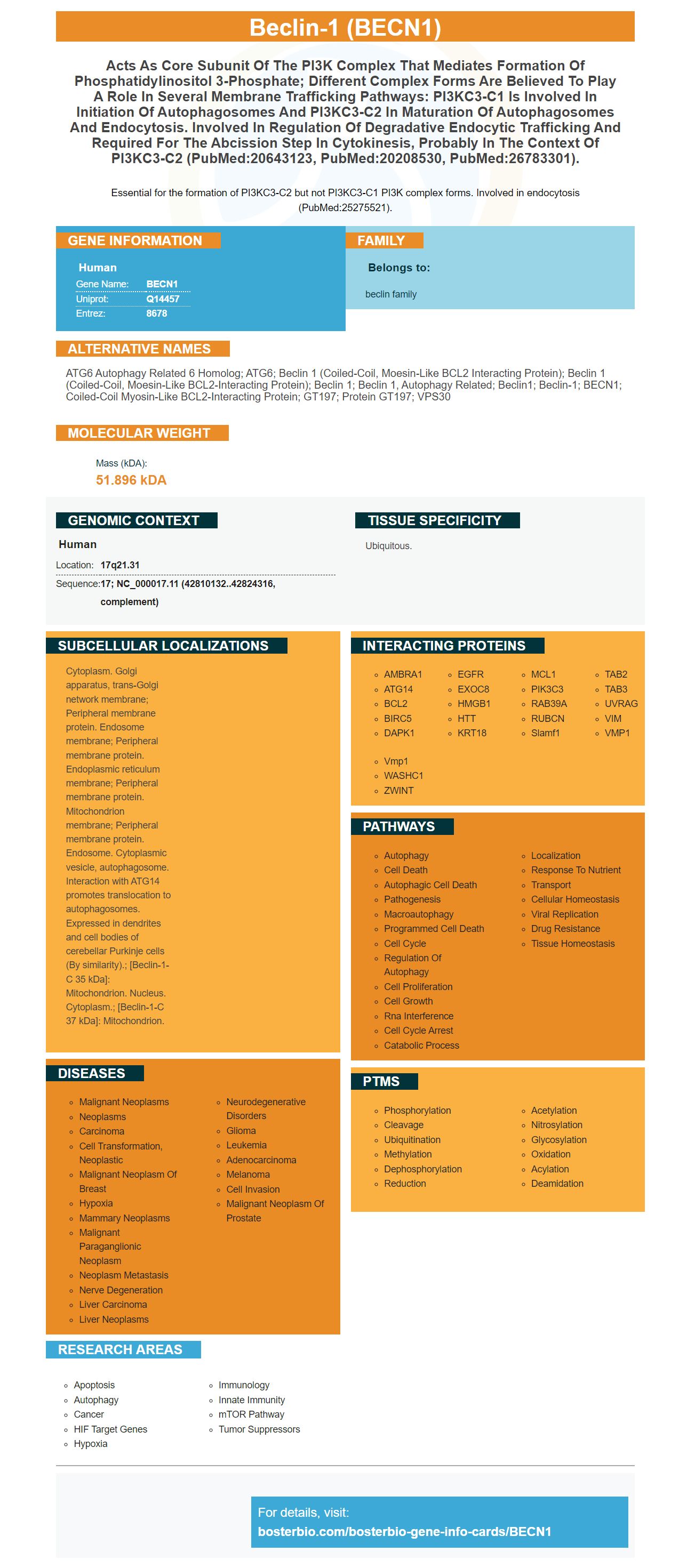
Facts about Beclin-1.
Essential for the formation of PI3KC3-C2 but not PI3KC3-C1 PI3K complex forms. Involved in endocytosis (PubMed:25275521).
Human | |
---|---|
Gene Name: | BECN1 |
Uniprot: | Q14457 |
Entrez: | 8678 |
Belongs to: |
---|
beclin family |
ATG6 autophagy related 6 homolog; ATG6; beclin 1 (coiled-coil, moesin-like BCL2 interacting protein); beclin 1 (coiled-coil, moesin-like BCL2-interacting protein); Beclin 1; beclin 1, autophagy related; beclin1; beclin-1; BECN1; Coiled-coil myosin-like BCL2-interacting protein; GT197; Protein GT197; VPS30
Mass (kDA):
51.896 kDA
Human | |
---|---|
Location: | 17q21.31 |
Sequence: | 17; NC_000017.11 (42810132..42824316, complement) |
Ubiquitous.
Cytoplasm. Golgi apparatus, trans-Golgi network membrane; Peripheral membrane protein. Endosome membrane; Peripheral membrane protein. Endoplasmic reticulum membrane; Peripheral membrane protein. Mitochondrion membrane; Peripheral membrane protein. Endosome. Cytoplasmic vesicle, autophagosome. Interaction with ATG14 promotes translocation to autophagosomes. Expressed in dendrites and cell bodies of cerebellar Purkinje cells (By similarity).; [Beclin-1-C 35 kDa]: Mitochondrion. Nucleus. Cytoplasm.; [Beclin-1-C 37 kDa]: Mitochondrion.
Boster Bio can be used to answer a variety of questions. Boster Bio's optimization manual can help you answer these questions and many more to improve your research. A troubleshooting guide will assist you in identifying and eliminating errors and ensure your experiment is as efficient as it can be. These guides also address common questions that researchers have about using Boster Bio. We'll talk about the most frequent issues with BECN1 as well as ways to make your experiments more efficient.
The ER contains more than half of the cell's bilayers made of lipids and is the source of lipids for the membranes of organelles. The ER lumen can trigger ferroptosis because of its oxygen-rich environment. However, the exact mechanism through which the ER starts ferroptosis is not clear. The mechanism is still not well understood but it is involved in autophagy and mitophagy.
A panel of epigenetic regulators are responsible for causing ferroptosis. The function of this panel of genes is to control gene transcription, cellular fate and development. These regulators are vital for ferroptosis and may be used as therapeutic targets. These findings are encouraging because they could be a way to remove an obstacle to treatment and prevention of cancer.
Autophagic suppression is also controlled by BECN1. Induced autophagy promotes ferroptosis by increasing the expression of BECN1. It is interesting to note that this process is regulated by AMPK which is a cell-based energy sensor. Furthermore, AMPK-mediated BECN1 phosphorylation is important for the formation of a pro-autophagic complex in the event of glucose starvation. AMPK Phosphorylation at S96 inhibits ferroptosis.
In addition to being a crucial regulator of ferroptosis BECN1 also regulates the expression of many other important proteins that impact the ER. In cancer the protein that controls ferroptotic cell death blocks the development of PDAC. BECN1 is vital for activation of the SLC7A11 gene and may be connected to autophagy.
Ferroptosis is caused by iron metabolism-related pathways and the redox reactions. Pre-autophagosomal structures can turn into autolysosomes. The fusion between autolysosomes promotes autophagy and subsequent degradation of their contents. This process is made easier by BECN1 and plays a key role in pancreatic cancer genesis.
Autophagy is a conserved pathway that removes cellular components via the Lysosomes. Autophagy functions are dependent on context and may either encourage or hinder tumor growth. Its role in ferroptosis is context-dependent but it is a necessary component of autophagy. Autophagy is a necessary component of ferroptosis. These pathways could be linked through a hidden pathway.
The BECN1 gene is involved in ferroptosis, is one of the main regulators of GPX4 activities. This enzyme transforms lipid hydrogen peroxide into an alcohol that is not toxic, a lipid alcohol, with the cofactor GSH and protects cells from lipid peroxides. This enzyme is inhibited by the inhibitor SystemXc- which also inhibits the synthesis of GSH and GPX4. Thus targeting the GPX4 enzyme could be an effective way to regulate ferroptosis.
GPX4 also plays a role in lipid peroxidation and knocking out the enzyme can lead to ferroptotic cell death. This enzyme also affects the iron levels and blocks the mevalonate pathway which, in turn, causes cell death. Additionally, GPX4 functions as an inhibitor of lipid peroxidation. Induction of GPX4 expression through the BECN1 gene inhibits ferroptosis and increases MAP1LC3B II production.
The BECN1 gene regulates the expression of xCT/GPX4 in cancer cells. In addition, BECN1 also inhibits lipid peroxidation. In addition, BECN1 is a member of the BRCA1 family. The expression of genes is controlled by the BRCA1-associated protein (BRCA1), the SLC7A1 subunit, and the BECN1 gene.
Besides, the BECN1 gene promotes the synthesis of ferrotene, a component that helps protect cells from iron-induced lipid peroxides. It is important to note that the BECN1 gene is not the only gene that regulates xCT/GPX4 expression. The BECN1 gene also plays an important role in the maintenance of normal iron levels in ferroptosis. It is a critical regulator for the health of all ferroptotic cells.
BECN1 is an important marker that has recently been identified as a potential therapeutic agent for fibrotic liver. In the present study, we have investigated the role of BECN1 in liver fibrosis. We used mice transgenic with the human BECN1 gene to examine the effect of a drug targeting BECN1 on liver fibrosis. The results showed that BECN1 knockdown prevented the development of liver fibrosis and inhibited MSC-ex-mediated ferroptosis.
BECN1 is a vital regulator of ferroptosis in HSCs. In addition to regulating ferroptosis, BECN1 regulates hepatic collagen deposition and is transported by MSC-ex. These results demonstrate the potential of BECN1 as an anti-fibrosis therapeutic. The drug's efficacy and safety is being tested in clinical trials.
The drug CCl4 and BDL induce liver fibrosis in mice. In addition, increased serum DDR1 levels are associated with LF. Further, LF levels are correlated with collagen I levels in the ECM of the liver. Immunohistochemistry reveals an increase in collagen deposition in patients with liver cirrhosis. But this relationship isn't clear yet.
It is thought that hypoxia regulates the expression of various genes, including those responsible for angiogenesis and collagen synthesis. Therefore, BECN1 is a promising therapeutic target for liver fibrosis. However, further research is required to confirm these findings. The company plans to submit a patent application in the near future. And if their application is successful, it could revolutionize hepatic fibrosis research.
BECN1 is an essential marker of autophagy, a cellular homeostatic mechanism to eliminate cytoplasmic toxic aggregates, damaged organelles and invading pathogens. In addition, abnormal autophagy has been implicated in various pathological conditions. Beclin1 is a Bcl-2 homology 3-domain protein, which assembles cofactors to form the BECN1-PIK3C3-PIK3R4 complex and triggers the autophagy protein cascade. Yet, its function in ES is unclear.
The BECN1 marker was originally isolated from human Ewing sarcoma, a rare and aggressive neoplasm that affects primarily adolescents and has a poor prognosis if it metastasizes. Using reverse transcription-PCR technology, researchers were able to screen 84 key autophagy genes for key regulators. The researchers identified heat shock protein 90 as a potential regulator and confirmed its role in autophagy/apoptosis.
The results of these studies suggest that BECN1 acts as a mediator between BCL2-BCLXL and ATG5. Furthermore, BCL2 family proteins may suppress autophagy by displacing BECN1. As a result, BECN1 and ATG5 function independently. The interaction between BECN1 and ATG5 has been found to be essential for efficient cell death response. The results of these experiments suggest that the anti-apoptotic effect of ATG5 is mediated by autophagy.
BECN1 expression levels were determined using human HAT-7 cells. In the study, BECN1 and mTOR were upregulated. Both mTOR and BeCN1 were upregulated in the NaF-induced autophagy group. However, BECN1 was not shown to inhibit the autophagy process. Furthermore, the BECN1 marker regulates autophagy in mammalian cells.
The results of this study also show that selenite decreases autophagy. This decreased autophagy may be due to the loss of binding of the BECN1 promoter. This result is in line with the results of other studies using selenite and 17-AAG as biomarkers. This work is an important step in developing cancer treatments. If successful, Boster Bio's BECN1 marker will provide useful information for clinical diagnosis.
In the study, 240 healthy ICR mice were divided into four groups. The control group received distilled water, while the experimental groups were given 12, 24, or 48 mg/kg NaF. The dose was administered intragastrically once daily for 42 days. The results revealed that NaF significantly reduced the levels of mTOR and PI3K/Akt. The study also showed that NaF inhibits mTOR activity, and increased the expression of LC3II.
PMID: 9765397 by Liang X.H., et al. Protection against fatal Sindbis virus encephalitis by beclin, a novel Bcl-2-interacting protein.
PMID: 10395800 by Aita V.M., et al. Cloning and genomic organization of beclin 1, a candidate tumor suppressor gene on chromosome 17q21.
*More publications can be found for each product on its corresponding product page