This website uses cookies to ensure you get the best experience on our website.
- Table of Contents
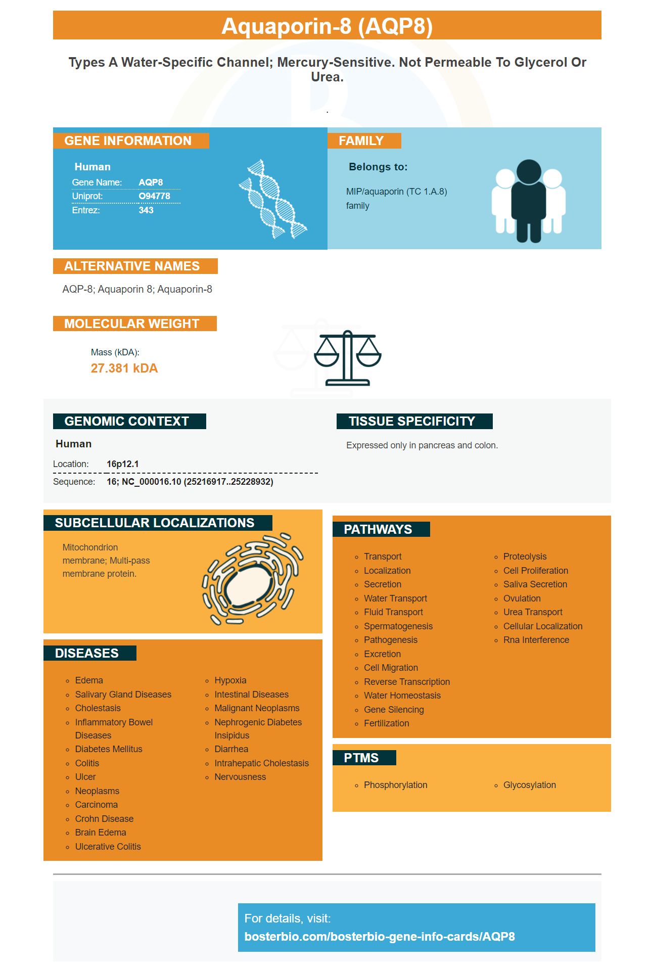
Facts about Aquaporin-8.
.
Human | |
---|---|
Gene Name: | AQP8 |
Uniprot: | O94778 |
Entrez: | 343 |
Belongs to: |
---|
MIP/aquaporin (TC 1.A.8) family |
AQP-8; aquaporin 8; aquaporin-8
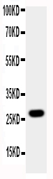
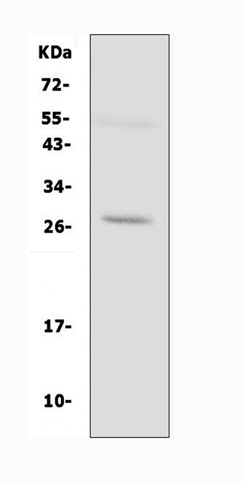
Mass (kDA):
27.381 kDA
Human | |
---|---|
Location: | 16p12.1 |
Sequence: | 16; NC_000016.10 (25216917..25228932) |
Expressed only in pancreas and colon.
Mitochondrion membrane; Multi-pass membrane protein.
This article will provide an overview of AQP8. Its best uses will be discussed with regard to applications and special samples. Boster scientists can submit their research for credit or special species. These product credit can be used worldwide to support scientific research. These are just a few examples of such applications. Read on to learn more! *Autoradiography film/Enhanced Chemiluminescence Detection (ECL).
The AQP8 marker was initially identified in cervical cancer tissues and normal cervix tissues. The AQP8 protein was strongly positive in cervical cancer tissues but non-reactive in adjacent normal samples. Its expression in different cell lines was determined. AQP8+ cells had higher IHC scores (mean) than normal cervical tissue. Western blot analysis in SiHa or HeLa cells revealed AQP8.
AQP8 comes from the aquaporin group of membrane proteins. Its main function involves the facilitation of small molecules, including water, being transported. AQP8 is implicated in many types of cancer, including esophageal. The exact mechanism by which AQP8 might contribute to cancer development remains unknown.
SiHa cells showed that AQP8 promoted cervical cancer metastasis. Western blot can also be used to detect classical EMT markers. E-cadherin and N-cadherin are both inhibited when AQP8 is expressed. Knockdown of AQP8 results in the reverse of EMT by restoring vimentin and N-cadherin expression.
AQP8 recognizes the mRNA for the human AQP8 gene in addition to the markers mentioned above. A cartoon representation was used to create a 3-D model for human AQP8 in a previous study. The cartoon representation represented amino acids in both the water selection and entrance regions as sticks. The sequence of human individual AQPs was consistent with the numbering of amino acids. Sequence identities between human AQP8 and the template model were highlighted in red while differences between AQP8 and the AQP5 marker were highlighted in light blue. The AQP8 Gene water channel is designated by W.
AQP8 was initially identified as a marker for a tumor cell line. Because it is highly conserved within its molecular landscape, the AQP8 mark is a candidate for this study. In addition, it was found to be essential in the transport of cell fluids. To date, thirteen AQPs can be identified. This gene was first cloned on October 28, 1998. It is involved in the regulation of cell fluid levels and characterized through comparative genomics.
Aquaporin 8 acts as a small, integral membrane protein and a water channel. Its gene encodes a sequence of amino acids. It is only found in pancreas. Six exons can be found in the Aquaporin-8 Gene. The gene maps directly to chromosome16p12. BosterBio has validated antibodies against Aquaporin 8
The AQP8 marker from Boster Bio has been validated on Western Blotting and Immunohistochemistry. It is highly sensitive and has good affinity. The antibodies can also be used in ELISA and Immunohistochemistry. AQP8 is a good choice for diagnostics as well as research. It is available from Boster Bio and is compatible to many biosensors.
ECL is a detection technology that uses antibodies conjugated HP. The enzyme reacts with the substrate to create excited intermediates that emit strong blue lights at 450nm. The light emission can be seen only during the enzyme/substrate react. Signal output stops when the substrate is depleted.
Researchers examined the effect of AQP8 upon cervical cancer cells by using transwell assays. Researchers discovered that SiHa cells expressing AQP8 migrated to invade more cells than the control cells. These abilities were reduced by knocking down the AQP8 genes. This study suggests that the AQP8 genes may be associated with higher cervical cancer cell viability.
The antibody failed detection of the molecule when it was absent AQP8. The 34-kDa band disappeared when the peptide had been preabsorbed with a molar surplus. After incubation, N-glycosidase was added to the peptide. The band shifted to its predicted 28kDa molecular mass. N-glycosylation means that the AQP8 molecule is present.
Western blotting can help detect proteins of particular interest. Colorimetric colorimetry uses secondary antibodies that have been conjugated to enzyme-reporters to produce a visually recognizable signal. These proteins are easily identifiable by the eye as distinct colors. Compared to other methods of protein detection, colorimetric detection is cheap and easy to set up.
Currently, colorimetric readouts are common in molecular diagnostics. Semi-quantitative analysis is possible. The detection level of AQP8 in the blood is sensitive enough to be used for a naked eye evaluation. One popular colorimetric system is the horseradish peroxidase H2O2 system. In this system, chromogen is linked to an enzyme and produces a colour by-product that signals the presence HDMRs.
PMID: 9806845 by Koyama N., et al. Cloning and functional expression of human aquaporin8 cDNA and analysis of its gene.
PMID: 28042826 by Laforenza U., et al. Aquaporin-Mediated Water and Hydrogen Peroxide Transport Is Involved in Normal Human Spermatozoa Functioning.