This website uses cookies to ensure you get the best experience on our website.
- Table of Contents
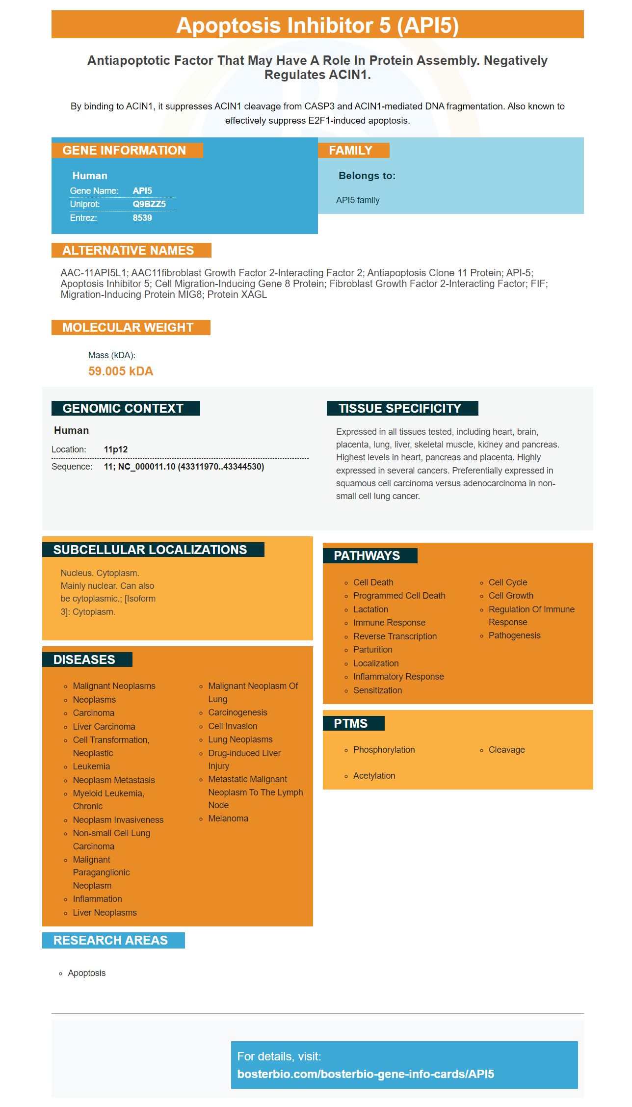
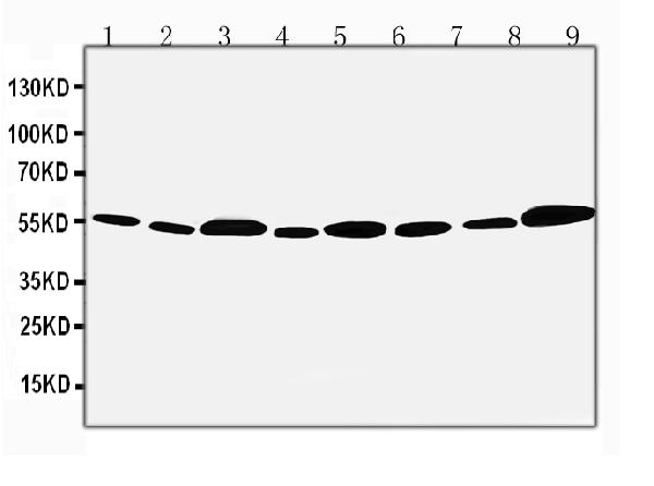
Facts about Apoptosis inhibitor 5.

By binding to ACIN1, it suppresses ACIN1 cleavage from CASP3 and ACIN1-mediated DNA fragmentation. Also known to effectively suppress E2F1-induced apoptosis.
| Human | |
|---|---|
| Gene Name: | API5 |
| Uniprot: | Q9BZZ5 |
| Entrez: | 8539 |

| Belongs to: |
|---|
| API5 family |

AAC-11API5L1; AAC11fibroblast growth factor 2-interacting factor 2; Antiapoptosis clone 11 protein; API-5; apoptosis inhibitor 5; Cell migration-inducing gene 8 protein; Fibroblast growth factor 2-interacting factor; FIF; migration-inducing protein MIG8; Protein XAGL
Mass (kDA):
59.005 kDA

| Human | |
|---|---|
| Location: | 11p12 |
| Sequence: | 11; NC_000011.10 (43311970..43344530) |
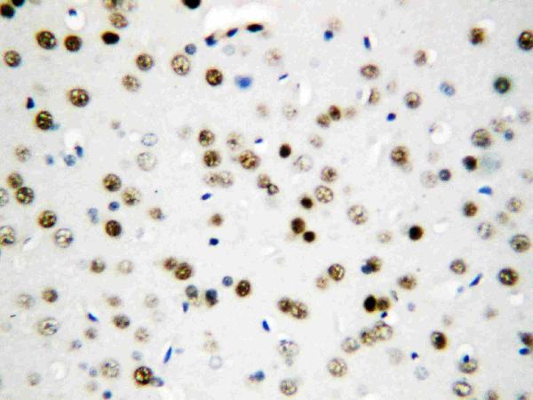
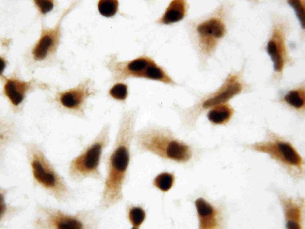
Expressed in all tissues tested, including heart, brain, placenta, lung, liver, skeletal muscle, kidney and pancreas. Highest levels in heart, pancreas and placenta. Highly expressed in several cancers. Preferentially expressed in squamous cell carcinoma versus adenocarcinoma in non-small cell lung cancer.
Nucleus. Cytoplasm. Mainly nuclear. Can also be cytoplasmic.; [Isoform 3]: Cytoplasm.


If you're in the market for a new anti-apoptosis antibody Boster Bio's Anti-Bax Monoclonal Antibody may be ideal for you. This monoclonal antibody targets Apoptosis-related Protein Bax, which is an essential marker for research. The API5 marker is a standard API used in the field of gene therapy, cell-culture studies and many other applications.
Monoclonal Antibody for Bax recognizes human Bax characterized by conformational change. This antibody recognizes the exact human Bax region in mouse, rat, or human cells. It is created by using an unionic detergent that exposes an epitope of Bax. The antibody is able to bind monomeric Bax but is not able to complex Bax with Bcl-2 and Bcl-xL.
The Anti-Bax Monoclonal Abortus marker is an anti-virus of high-quality that has been extensively tested in IHC and WB. It reacts with Monkey and Human cells. It also contains BSA as a preservative. It is suitable for a variety of immunological research objectives.
The Anti-Bax monoclonal antibody can be compatible with other mAbs. Anti-Bax antibody is a good choice in studies where Bax and Bcl2 are both correlative. It can be stored at 2-8 degC for up to one week. Aliquots are best stored at -20degC. Antibody solutions should be mixed gently before use.
The key process in the field of cell biology is detection of Bax. In vitro research has demonstrated that bax is essential for apoptosis, although its role is still unclear. Monoclonal antibodies against Bax can detect Bax, and inhibit apoptosis. These antibodies are able to detect Bax and its Oligomerization.
Flow Cytometry has been used to determine Bax in human cells. HCT116 cells that express GFP-Bax as well as a mutant form were exposed to 25 mm of H2O2 for one hour. The cells were then transfected using anti-GFP-Bax antibodies. After the transfection, the cells were subjected to AMS and SDS-PAGE. The anti-Bax antibody was used as a protein loading control.
The Cys-Bax mutant C126S/K64A and GFP-Bax C126S/K57A/K58A had no on the subcellular distribution of Bax. However, they did result in the activation of Bax. This mutant form of Bax is sensitive to H2O2 in Bax deficient HCT116 cells. In addition, it does not change the H2O2-dependent activity of Cys-62.
Bak Ab-1 is a protein that binds to Bak and has immunofluorescence associated with it. It is also linked to Apoptotic and pre-apoptotic cells. Both antibodies recognize the same epitope in cells, as observed in Fig. 5 comparison analysis. However the antigen polyclonal to Bak has a higher level of fluorescence correlated with the treatment of etoposide.
The manufacturer of the antibody guarantees the product for one year. The antibody was tested against a variety of types of cancer cells in mouse human, human, and rat tissue. Its effectiveness has been confirmed through clinical trials. The World Health Organization (WHO), has recommended it as an extremely sensitive antibody for diagnose cancer using cells. It has a wide array of uses.
Flow cytometric analysis of antibody's function is performed in two different cell lines. The two cell lines used in these tests were 293T cells that were challenged with SVV at different MOIs. The cells were harvested at the specified times and stained with Annexin V-FITC/PI and observed under a fluorescence microscope.
The Anti-Bax (Apoptoses Marker) monoclonal antibody of the Boster Bio is an antigen receptor with chimeric structure that detects Bax the apoptotic protein. This antibody was evaluated using an xenograft from mice that had tumors. The tumor was pulverized through a 100-mm cell strainer . It was then was lysed in RIPA buffer using a harsh pipetting. Secondary antibodies were employed against Bax, C-actin and b-actin.
The Anti-Bax (Apoptoses Marker) Monoclonal antibody from Boster Bio has been validated using ELISA as well as WB and IHC methods. It reacts with Human, Mouse, and Rat. It is well-suited for studies on Apoptosis marker protein. Antibodies from Boster Bio are designed to recognize markers of apoptosis and to cause cell death by inhibiting the activity of apoptosis.
This immunotherapy target is derived by mimotopes that originate from the immune checkpoints Bax and TNF-alpha. These may lead to the development of cancer vaccines. This discovery may also pave the way for therapies that combine tumor-specific antigens. This new approach could increase the effectiveness of vaccinations as well as cancer vaccines. It can also be adapted to the type of tumor and its development stage.
Rabbits are the main source of the anti-Bax antibody. The antigen BHRF1 was administered to the rabbit. The mice that responded the most quickly were sacrificed. Tsai and colleagues have described the process of creating hybridoma cells. (58). By using immunofluorescence tests and Western blot analysis the antibody clone that had the highest reactivity to BHRF1, 3E8 was chosen. The specificity of the cloned antibody was determined.
PMID: 9307294 by Tewari M., et al. AAC-11, a novel cDNA that inhibits apoptosis after growth factor withdrawal.
PMID: 10393420 by Gianfrancesco F., et al. Molecular cloning and fine mapping of API5L1, a novel human gene strongly related to an antiapoptotic gene.