This website uses cookies to ensure you get the best experience on our website.
- Table of Contents
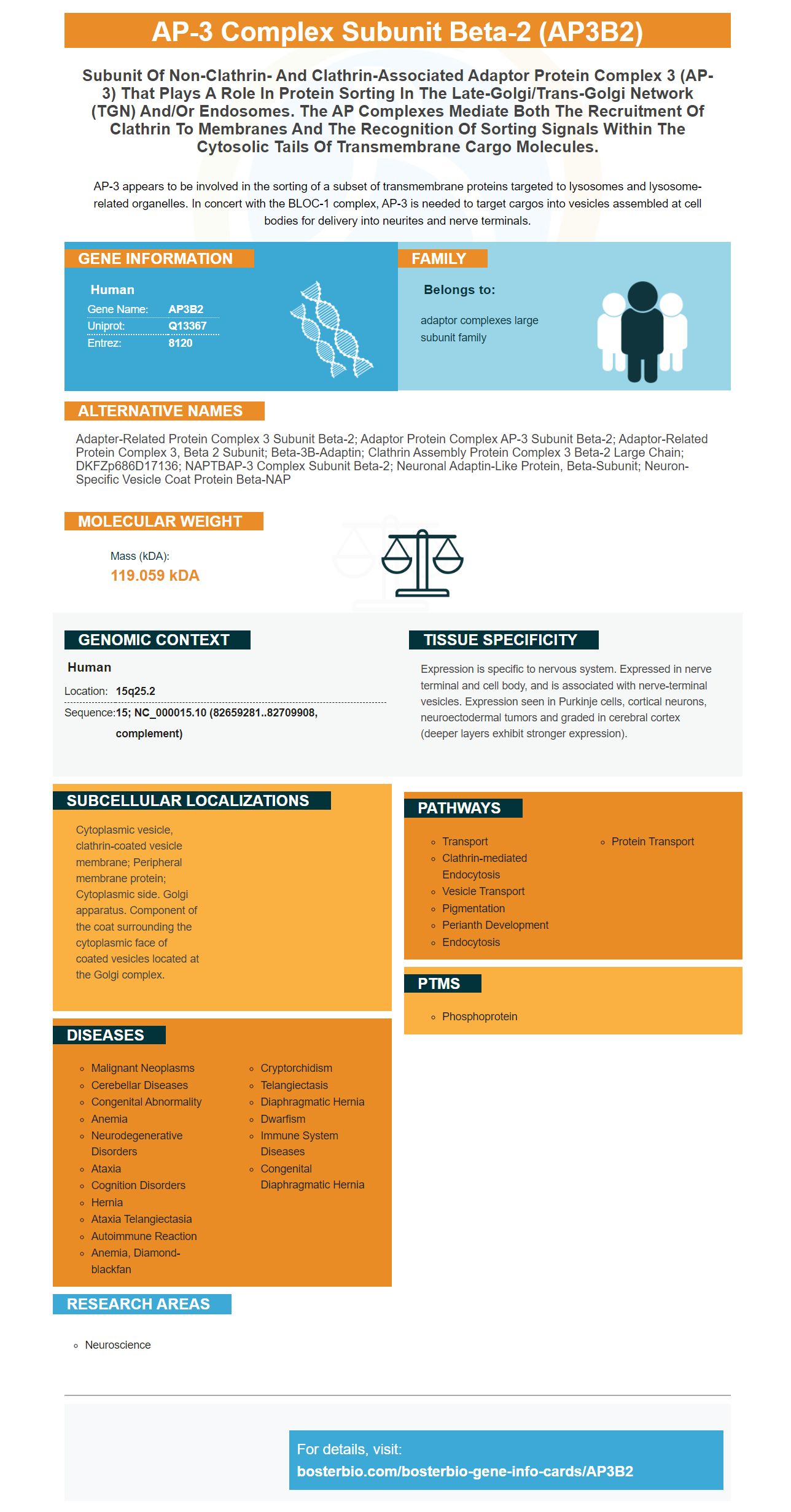
Facts about AP-3 complex subunit beta-2.
AP-3 appears to be involved in the sorting of a subset of transmembrane proteins targeted to lysosomes and lysosome-related organelles. In concert with the BLOC-1 complex, AP-3 is needed to target cargos into vesicles assembled at cell bodies for delivery into neurites and nerve terminals.
Human | |
---|---|
Gene Name: | AP3B2 |
Uniprot: | Q13367 |
Entrez: | 8120 |
Belongs to: |
---|
adaptor complexes large subunit family |
Adapter-related protein complex 3 subunit beta-2; Adaptor protein complex AP-3 subunit beta-2; adaptor-related protein complex 3, beta 2 subunit; beta-3B-adaptin; Clathrin assembly protein complex 3 beta-2 large chain; DKFZp686D17136; NAPTBAP-3 complex subunit beta-2; Neuronal adaptin-like protein, beta-subunit; Neuron-specific vesicle coat protein beta-NAP
Mass (kDA):
119.059 kDA
Human | |
---|---|
Location: | 15q25.2 |
Sequence: | 15; NC_000015.10 (82659281..82709908, complement) |
Expression is specific to nervous system. Expressed in nerve terminal and cell body, and is associated with nerve-terminal vesicles. Expression seen in Purkinje cells, cortical neurons, neuroectodermal tumors and graded in cerebral cortex (deeper layers exhibit stronger expression).
Cytoplasmic vesicle, clathrin-coated vesicle membrane; Peripheral membrane protein; Cytoplasmic side. Golgi apparatus. Component of the coat surrounding the cytoplasmic face of coated vesicles located at the Golgi complex.
This article provides information about the Boster Biologics marker and the AP3B2 marker. This article will examine Steven Boster as well as the AP3B2 mark. We will also discuss the differences between rabbit and mouse and what we can do to use the marker in our experiments. This article will help you get started with your experiments with the AP3B2 markers.
The AP3B2 Gene is one the most used proteins in research and diagnostics. Many biological tests use antibodies to detect AP3B2 within animal samples. Boster Bio uses rabbit and mouse models to develop antibodies to the AP3B2 Gene. Although this gene is widely available, not many companies have antibodies that are specifically designed for it. For this reason, it's essential to perform your own experiments with the antibodies.
AP3B2 can be used to identify the antigen apolipoprotein B. Many biological assays detect AP3B2 using antibodies. The antibodies can be monoclonal or polyclonal, and have broad spectrum binding capabilities that allow them to detect AP3B2 from many animal samples. Boster bio.com uses rabbit and mouse models to develop its AP3B2 antibody.
The lysosomal protein AP3B2 is the source of the AP3B2 marker. It is essential for endosomal trafficking cargo to the lymphosomes. The AP3B1 subunit was associated with the hermansky–pudlak condition, which is a genetic condition characterized in part by lysosomal LRO defects and albinism. The AP-3 Complex includes many subunits which are tissue-selective, differentially expressed, and tissue-selective.
Standard methods were used to genotype mice by PCR. The AP3B2 genetic code was expressed in a variety o tissues, including the heart, liver, and kidney. Genotyping was performed by using DNA extracted from tails and primers specific for Rab7l1 wild-type allele and Lrrk2 wild-type allele. Mice were fixed in 4% paraformaldehyde and perfused for a minimum of 24 hours.
AP3B2 antibodies are produced by boster bio and were originally demonstrated to colocalize with patient IgG on mouse brain tissue. Boster bio developed the antibodies with the help of immunohistochemistry, which detected AP3B2 antibodies in human sera and recombinant AP3B2 polypeptide. The sera of patients were compared to those from healthy individuals and the control groups. The results in the AP3B2-specific cells-binding assay were favorable.
The AP3B2 antibody produced the same staining pattern in both CSF and serum samples. These patterns were predominant in the cerebellum and in spinal cord gray material, but they could also be seen in peripheral sensory ganglia and sympathetic ganglia. CBA, western blot and other evidence show that AP3B2 immunodeficiency is associated also with a symptomatic neurologic condition. 9 patients with subacute and rapidly progressing gait ataxia were studied. Myeloneuropathy, peripheral sensor neuropathy, cerebellar, cerebellar, and spinocerebellar symptoms were some of the neurologic manifestations.
Antibodies against AP3B2 are used in biological tests to detect the protein within various organisms. Antibodies come in monoclonal as well as polyclonal forms and can react with AP3B2 from a variety of animal specimens. Boster Bio also offers antibodies that can be used in research on neurologic disorders and the study neuromuscular diseases.
Online immunohistochemical images of 12 reactivities for Boster Bio AP3B2 can be viewed. They are validated using known positive and negative samples. All current assays are reviewed. Typical high-resolution immunohistochemical images are presented. Diagnostic pitfalls are discussed and all available assays are reviewed. In addition to the Boster Bio AP3B2 antibody, a boster immunohistochemical analysis of human hepatic tissue will confirm the presence of a positive AP3B2 protein.
PMID: 7671305 by Newman L.S., et al. Beta-NAP, a cerebellar degeneration antigen, is a neuron-specific vesicle coat protein.
PMID: 17453999 by Chen C., et al. Characterization of AP3B2_v2, a novel splice variant of human AP3B2.