This website uses cookies to ensure you get the best experience on our website.
- Table of Contents
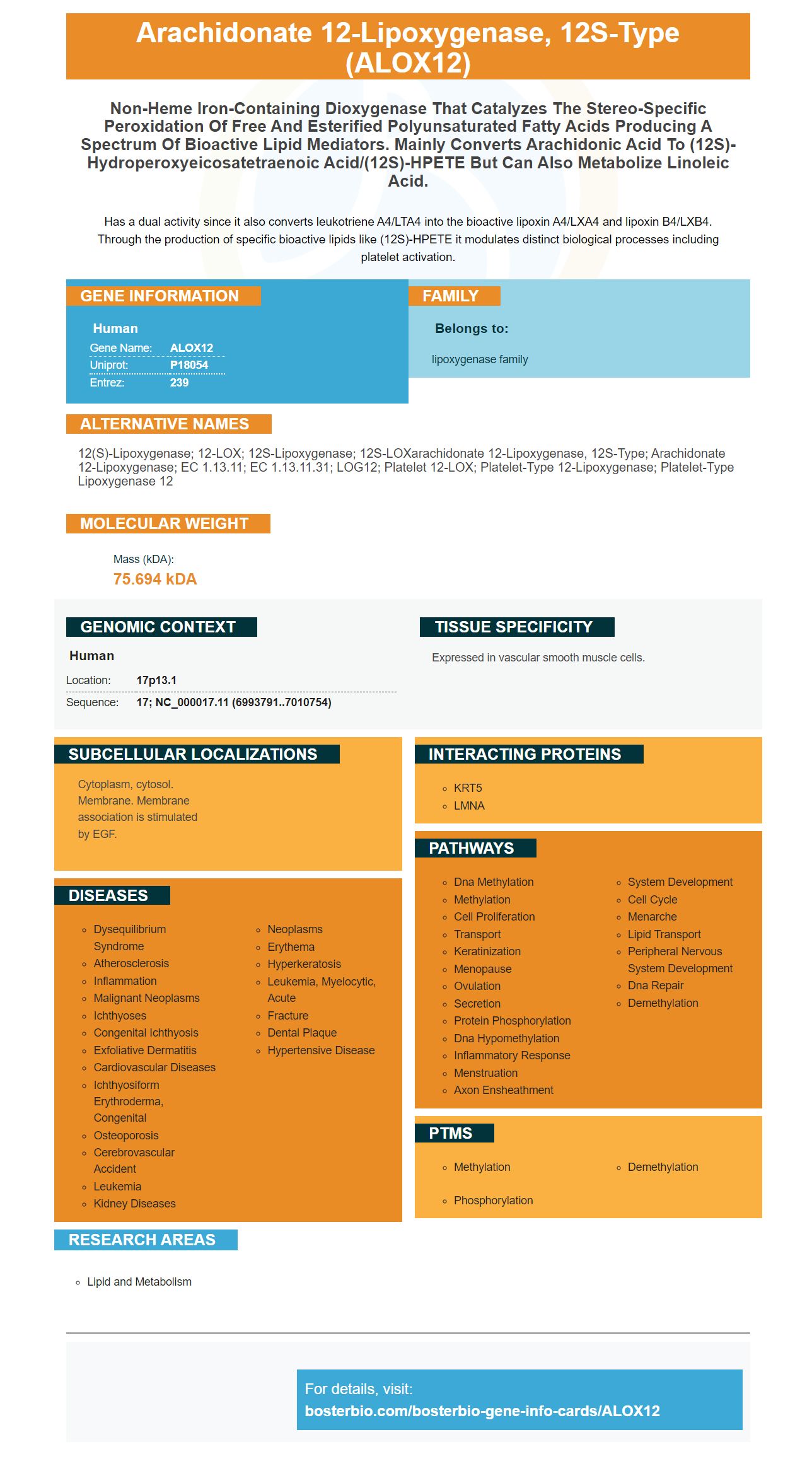
Facts about Arachidonate 12-lipoxygenase, 12S-type.
Has a dual activity since it also converts leukotriene A4/LTA4 into the bioactive lipoxin A4/LXA4 and lipoxin B4/LXB4. Through the production of specific bioactive lipids like (12S)-HPETE it modulates distinct biological processes including platelet activation.
Human | |
---|---|
Gene Name: | ALOX12 |
Uniprot: | P18054 |
Entrez: | 239 |
Belongs to: |
---|
lipoxygenase family |
12(S)-lipoxygenase; 12-LOX; 12S-lipoxygenase; 12S-LOXarachidonate 12-lipoxygenase, 12S-type; arachidonate 12-lipoxygenase; EC 1.13.11; EC 1.13.11.31; LOG12; platelet 12-LOX; platelet-type 12-lipoxygenase; Platelet-type lipoxygenase 12
Mass (kDA):
75.694 kDA
Human | |
---|---|
Location: | 17p13.1 |
Sequence: | 17; NC_000017.11 (6993791..7010754) |
Expressed in vascular smooth muscle cells.
Cytoplasm, cytosol. Membrane. Membrane association is stimulated by EGF.
Anti-12 Lipoxygenase/ALOX12 Marker has many uses in human biology. It may facilitate ferroptosis. Listed below are some of the best uses of this marker for human biology. If you're a scientist looking for a new antibody to detect this marker, Boster has you covered. The antibody is validated on multiple platforms using known positive and negative samples. Boster rewards its first reviewers with product credits, and thanks all scientists around the world for their support.
The Arachidonate 12-lipoxygenase (ALOX12R) is a cytochrome P450 enzyme that metabolizes arachidonic acid to 12(R)-HpETE. The cytochrome P450 enzymes convert arachidonic acid to various derivatives, including 12(S)-HpETE. The Anti-12 Lipoxygenase/ALOX12 Marker is a specific antibody that binds to human ALOX12R.
The enzyme is an essential component of the metabolic syndrome, a set of factors that can contribute to cardiovascular disease, diabetes, and obesity. In humans, it metabolizes arachidonic acid to 15(S)-HpETE, while in mice, it metabolizes arachidnic acid to 12(S)-HpETE.
AA 12-lipoxygenating ALOX15 is the most common ortholog in mammals. In humans, it is classified as an AA 15-lipoxygenase, while its ortholog in brown rats and house mice is AA 12-lipoxygenase. These two isoforms coexist, but they are not randomly distributed among mammals.
The Triad Concept was developed based on mutagenesis studies of selected ALOX15 orthologs. This model can be used to predict reaction specificity in mammalian ALOX12 orthologs without functional data. However, this model does not explain the AA 15-lipoxygenating ALOX15B. And it is unclear whether the anti-AA 15-lipoxygenase marker is specific for ALOX15B.
The Anti-12 Lipoxygenase/ALOX12 Marker is used to determine whether an ALOX12-containing protein is present in a cell. This enzyme is expressed in a variety of tissues including skin, heart, and kidney. Boster Bio's Anti-12 Lipoxygenase/ALOX12 Marker is available in multiple forms.
CYP1A1 is the main enzyme responsible for LPS-induced inflammation in macrophages. However, its role in the immune response remains unclear. Despite this, studies have shown that overexpression of the 12-lipoxygenase gene enhanced IL-6 and TNFa production in RAW cells. Interestingly, 12S-HETE and TNFg were associated with decreased beta-cell secretion in mice that lacked the enzyme.
The cDNA sequence of the anti-12 Lipoxygenase/AOLX12 Marker is available in the market. Moreover, this antibody can be used as a reference in various researches relating to the biochemical processes in plants. These enzymes are important in the regulation of many vital biological processes. They have been extensively studied and are essential in plant stress tolerance.
The ALOX15 orthologs of the human AA 12 lipoxygenase were identified in two different taxons, the bamboo rat and the siamang. In addition, the ALOX15 ortholog of the mouse expressed the Phe353-Ile418-Val419 motif. Both the collared anteater and the giant anteater express ALOX15 orthologs.
As AA-OOH-PE levels increase in the body, it is believed that selenium proteins and ACSL4 facilitate ferroptosis. The two proteins do not only regulate lipid peroxide levels, but also participate in other physiological processes. Although the roles of these enzymes are not entirely understood, it is suspected that they may play a role in T2DM pathogenesis. This is a potential clinical benefit of ACSL4 for the treatment of type 2 diabetes.
Ferroptosis is a form of cell death induced by the accumulation of iron-dependent lipid peroxides. Cells undergo the cellular apoptotic response to excessive AA-OOH-PE. There are two types of small-molecule inducers of ferroptosis: those that inhibit system X C or glutathione peroxidase 4 (GPx4), and those that block specific sulphur-transfer pathways.
Ferroptosis is a process in which catalytic iron and thiols interact. This process occurs in the body but is rare in cancer development. While it is still largely unknown what the molecular mechanisms underlying ferroptosis are, many cancer cells display evidence of this process. In addition to facilitating ferroptosis, pro-ferroptotic treatments may be beneficial in the treatment of cancer.
Inducers of ferroptosis include BI-6c9 and elastin. Both BI-6c9 and elastin exhibit the characteristic of intrinsic disorder that allows them to participate in promiscuous regulation and interaction. These proteins are found in many cell types, including apoptosis, autophagy, and necroptosis. Inducers of ferroptosis are also capable of selectively inducing cell death.
Since its discovery, ferroptosis has been studied in various pathogenic processes. Some studies focus on disease related to kidney cells, immune cells, and neurons. Ferroptosis may also play a role in the development of certain types of diabetes. And since ferroptosis is an integral part of the progression of diabetes, it is important to monitor the effects of iron on the pancreas.
Interestingly, some cancer treatments target the SLC7A11 gene, which regulates glutamate and cystine transport. The system xc-subunit regulates the glutathione content of the intracellular compartment while facilitating the transport of glutamate and cystine into the cell. However, not all P53 transcriptional targets facilitate ferroptosis. In other words, some drugs inhibit the sulphur-transfer pathways to decrease cystine and glutamate import, which facilitate ferroptosis.
Recent studies indicate that NCOA4 promotes ferroptosis by promoting ferritinophagy. The enzyme binds ferritin, allowing the iron to be released. The iron is then delivered to the lysosomes for degradation, resulting in an increase in the cell's iron level. However, this process depends on the P53-P21 axis and may further enhance ferroptosis.
The role of Haem oxygenase-1 in ferroptosis is unclear, but this enzyme plays an important role in maintaining the balance of intracellular iron. Ferroptosis inhibits cancer cells by reducing ROS levels. This, in turn, disrupts the balance between oxidation and reduction in cells. This may be why erastin has such a profound effect on lung cancer cells.
While the mechanism of GSH-mediated ferroptosis is not completely understood, some evidence suggests that it may be a key regulator in the cellular oxidation-reduction process. GSH mediates the reduction of lipid peroxides and organic hydroperoxide products in cells, enabling GPx4 to play a central regulatory role in ferroptosis. Moreover, it is known that cystathionine-b-synthetase activates methionine-to-cysteine conversion and promotes GSH synthesis through sulphur-transfer pathways. However, loss of this enzyme inhibits erastin-induced ferroptosis.
PMID: 2244907 by Yoshimoto T., et al. Molecular cloning and expression of human arachidonate 12- lipoxygenase.
PMID: 2377602 by Funk C.D., et al. Molecular cloning, primary structure, and expression of the human platelet/erythroleukemia cell 12-lipoxygenase.