This website uses cookies to ensure you get the best experience on our website.
- Table of Contents
2 Citations 8 Q&As

Facts about A disintegrin and metalloproteinase with thrombospondin motifs 1.

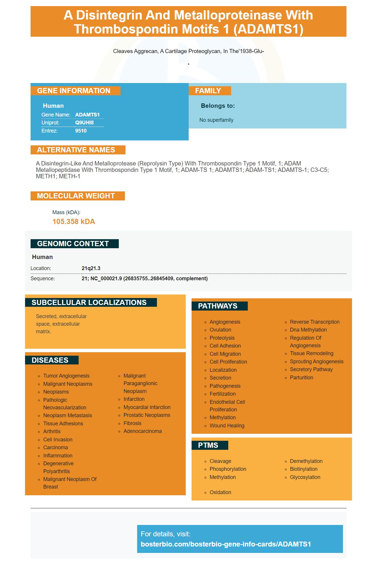
Cleaves aggrecan, a cartilage proteoglycan, in the'1938-Glu-
.| Human | |
|---|---|
| Gene Name: | ADAMTS1 |
| Uniprot: | Q9UHI8 |
| Entrez: | 9510 |

| Belongs to: |
|---|
| No superfamily |

a disintegrin-like and metalloprotease (reprolysin type) with thrombospondin type 1 motif, 1; ADAM metallopeptidase with thrombospondin type 1 motif, 1; ADAM-TS 1; ADAMTS1; ADAM-TS1; ADAMTS-1; C3-C5; METH1; METH-1
Mass (kDA):
105.358 kDA

| Human | |
|---|---|
| Location: | 21q21.3 |
| Sequence: | 21; NC_000021.9 (26835755..26845409, complement) |
Secreted, extracellular space, extracellular matrix.


ADAMTS1 is an expression gene found in many cells and tissues, including oocytes, primary granulosa cells(PGC), human malignancies and conditioning medium. It has many possible uses and researchers are looking to discover new applications. This article will go over its applications in these situations. Continue reading to learn more about ADAMTS1.
ADAMTS1 is a cytokine that regulates the quality and quantity of oocytes. Studies have shown that ADAMTS1's deregulation could cause a decline in the development of embryos. Researchers have discovered that ADAMTS1 reduces the levels of PTX3 and PTX4 in oocytes. This protein may impact the quality of oocytes and embryos, as well as the follicular microenvironment for PCOS patients.
The ovary's granulosa cell makes ADAMTS1 in its granulosa. Mice with this gene do not also subfertile and have fewer healthy-growing follicles. In women who are ADAMTS1 deficient, ovulation is affected and 45 percent of the newborn mice die due to renal malformation.
To study the role played by these proteins in embryonic development using animal models, we tested that had ADAMTS1 and ADAMTS5 silenced. The underlying cause of reduced ADAMTS1 expression could be due to hormones or paracrine elements that are located in the testis. Furthermore, the interaction between autocrine and paracrine hormones, temperature and blood supply could affect the expression of ADAMTS proteins.
This study investigated the effects of siRNA on ADAMTS-1 expression in human endothelial cells through transfection with a mock RNA (siRNA). The conditioned medium of cells transfected using scrambled siRNAs functioned as controls. Immunoblot analysis revealed a band with 80 kDa in the conditioned medium and 40 kDa in cell lysates. These results prove that siRNA-mediated knockdown works for ADAMTS-1.
Mutations in ADAMTS-1 have been discovered that disrupt the zinc binding motif. The X5(E386Q) mutant, which was expressed in COS-7 cells, did not contain high-molecular-weight species and lost binding capacity to a2M. Mutants of X5(E386) were not able to form zinc-binding compounds with a2M. However, the processed protein was released at levels similar to the X5.
The transfection of one ADAMTS-1 vector into a COS-7 line results in a doublet amount X4 proteins in the supernatant. However, cotransfection of mice's furin cDNA in LoVo cells result in a single band of ADAMTS-1 protein that has been processed. The result of this experiment suggests that the ADAMTS-1 X5 precursor can be cleaved at a single site by a furin endopeptidase vivo.
Researchers have found a lower amount of ADAMTS-1 in colon cancer cells than normal lung tissue in 1997. The decreased expression was linked with the transition from normal epithelial cells to cancer cells. While this may appear surprising, this finding could be significant in many ways. We will discuss the significance of this finding and what it means for the future the treatment of colon cancer patients.
While there isn't any concrete evidence linking ADAMTS-1 expression to cancer cells, researchers did observe that the mRNA levels of ADAM-12 were elevated in tumors. While ADAM-12 and ADAMTS-1 mRNA levels increased in cancerous cells, they decreased in healthy tissues. The authors also found a positive correlation between the mRNA levels of the two proteins and VEGF A121 expression in tumour cells.
The ADAMTS-1 gene is overexpressed in a variety of human malignancies, including breast cancer. It is believed that this overexpression causes cancer by altering the behavior and properties of tumor cells. This could, in turn cause the development of distant metastases. The results suggest a role for ADAMTS1 in malignancies of the human body, and the study provides further evidence of the significance of the ADAMTS1.
We used immunofluorescence to stain the expression of ADAMTS-1 in primary cells. Primary granulosa cells are cultivated in 6-well plates. They were then cultured for 4-5 days prior to transfection. At 6 h post-transfection cells were incubated for 6 h with testosterone (10-7 mol/L) in PBS. After washing the cells two times with PBS the cells were then probed for the ADAMTS-1 and Ki-67 proteins with an anti-rat secondary antibody. The Zeiss microscope was used to carry out the DAPI staining.
The ADAMTS-1 gene has been implicated in the regulation of numerous genes involved with follicular growth. In addition, down-regulation of ADAMTS1 inhibits the secretion of estrogen in granulosa cells. Additionally, it is believed to affect the expression of several genes in the embryonic stage that include the HLA-G gene, PAF, and LIF. This discovery has significant implications for the development of oocytes.
ADAMTS1 is involved in the ovulation of murine oocytes as well as in the structural remodeling of follicle borders of the stromal. However, its role in the development of embryos and oocyte quality is still unclear. However it has been established that ADAMTS1 expression increases in the granulosa cells of PCOS patients. Silencing ADAMTS1 can inhibit cell proliferation and the release of E2, a key factor in the embryonic development process. This could be due reductions in Bcl2 gene family genes.
Protrusions of the membrane that are invading are a characteristic of metastatic cancer cells invadopodia. These protrusions can penetrate the vasculature as attachment points. Recent studies have provided new insight into the networks of signaling that regulate invadopodia formation. These studies have identified potential targets for new therapies for metastatic disease.
The effect of ADAMTS-1 knockdown on invasion was observed in MCF7 cells, which is a noninvasive breast cancer cell line. When silenced in the cells ADAMTS-1 knockdown slowed down the rate of cell migration in MCF7 cells. The knockdown of ADAMTS-1 had no effect on the rate of migration in other cell types. This suggests that ADAMTS-1 could regulate invadopodia formation through direct regulation.
We transfected siRNAs into MDAMB-231 cells to find out if knockdown of ADAMTS-1 enhances invadopodia production. To determine the levels of their expression we used siRNAs containing scrambled sequences as control. Then, we used the conditioned media from these cells to induce tubulogenesis in human endothelial cells.
In a recent study, researchers identified that ADAMTS-1 expression is significantly regulated in the GCs in PCOS patients when compared to nonmovulatory controls. These results show that ADAMTS-1 expression is affected by dysregulation. These results confirm previous findings that ADAMTS1 could play a role in regulating oocyte maturity and quality.
ADAMTS-1 regulates several genes involved in embryonic growth and oocyte maturation. The silence of ADAMTS1 may increase the expression of genes associated with the process of apoptosis. After silence, Bcl-2 as well as Bcl-XL are increased. Additionally, oocytes created by mice who are deficient in ADAMTS1 have higher oocyte maturation rates and embryos of high-quality.
The study also revealed that ADAMTS-1 was not negatively related to D3 and D5 embryos of high quality in PCOS patients. Despite the small sample size the results suggest that ADAMTS1 may play a protective role in the development of Oocytes. In addition, the down-regulation of ADAMTS1 could decrease the number of oocytes that develop into good-quality embryos.
In the PCOS patients, ADAMTS-1 is expressed in the granulosa cells, which could indicate an ineffective ovulatory signaling. These findings will provide new information about the follicular microenvironment and embryo development in PCOS patients. It is important to remember that further research needs be carried out before conclusions can be drawn.
The discovery of Steven Boster's ADMTS1, marker for ovarian cancer , has prompted researchers to investigate whether the gene is involved in the function of ovarian. This protein affects mitochondrial pathway and the ratio of Bcl2 to Bax. If the gene is involved in the function of the ovary, it could cause premature ovarian failure, and the growth of ovarian cancer.
ADAMTS1 expression is variable in the early embryonic growth. In pre-implantation embryonic development, ADAMTS1 staining is mostly found in the cytoplasm. As embryos develop, staining intensity decreases. Interestingly, silencing ADAMTS1 significantly extended the time to the morula stage but did not affect the number of embryos that are good quality. The statistical significance was determined at 0.05.
PMID: 10438512 by Vazquez F., et al. METH-1, a human ortholog of ADAMTS-1, and METH-2 are members of a new family of proteins with angio-inhibitory activity.
PMID: 10785405 by Glienke J., et al. Differential gene expression by endothelial cells in distinct angiogenic states.
*More publications can be found for each product on its corresponding product page


