This website uses cookies to ensure you get the best experience on our website.
- Table of Contents
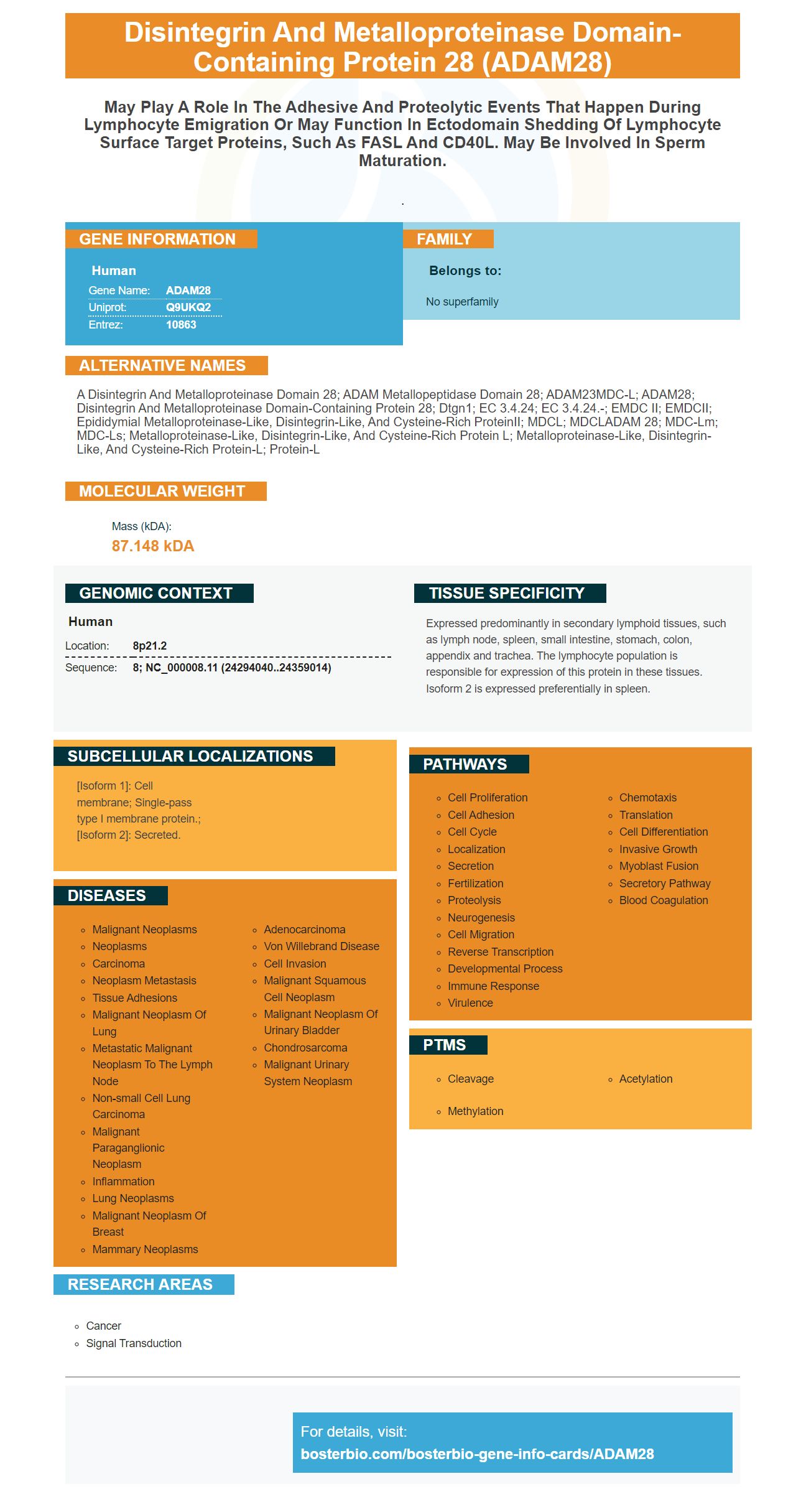
Facts about Disintegrin and metalloproteinase domain-containing protein 28.

.
| Human | |
|---|---|
| Gene Name: | ADAM28 |
| Uniprot: | Q9UKQ2 |
| Entrez: | 10863 |

| Belongs to: |
|---|
| No superfamily |

a disintegrin and metalloproteinase domain 28; ADAM metallopeptidase domain 28; ADAM23MDC-L; ADAM28; disintegrin and metalloproteinase domain-containing protein 28; Dtgn1; EC 3.4.24; EC 3.4.24.-; eMDC II; eMDCII; Epididymial metalloproteinase-like, disintegrin-like, and cysteine-rich proteinII; MDCL; MDCLADAM 28; MDC-Lm; MDC-Ls; Metalloproteinase-like, disintegrin-like, and cysteine-rich protein L; metalloproteinase-like, disintegrin-like, and cysteine-rich protein-L; Protein-L
Mass (kDA):
87.148 kDA

| Human | |
|---|---|
| Location: | 8p21.2 |
| Sequence: | 8; NC_000008.11 (24294040..24359014) |
Expressed predominantly in secondary lymphoid tissues, such as lymph node, spleen, small intestine, stomach, colon, appendix and trachea. The lymphocyte population is responsible for expression of this protein in these tissues. Isoform 2 is expressed preferentially in spleen.
[Isoform 1]: Cell membrane; Single-pass type I membrane protein.; [Isoform 2]: Secreted.


Antibodies against the ADAM28 marker may prove useful when working with cells from human beings. It is found in both whole-cell as well as cell-free samples. These antibodies react to ADAM28 DNA from rats, mice, and humans and are available in Boster Bio Catalog number A06873.
The ADAM28 marker plays an important role in the process. Researchers have found that this gene is crucial for the development Alzheimer's disease. The antibody to ADAM28, a boster bio-designed antigen that reacts with both Human proteins and Mouse proteins, is called a boster biodesigned antibody. This antibody can be used for several different applications, including the study of aging, cancer, and brain development.
It is still not known what the ADAM28 markers are best used for. Some studies suggest that ADAM28 may play an important function in sperm maturation, immune suppression, and other processes. ADAM28 expression has been found in the epididymis of 34% and 34% respectively in patients with lung cancer. However, the exact tissue distribution of the ADAM28 protein in normal human organs remains elusive.
Numerous studies have shown that ADAM28 is expressed by epithelial cell populations of normal organs including the bronchioles. It has been shown to affect cell survival and decrease cytotoxicity from the C1q enhancer. Similar to C1q, ADAM28 was found to inhibit C1q activity in bronchial epithelial cell lines. It is believed to play an important role in the development and progression of lung diseases.
The human genome hosts the ADAM28 gene. An ADAM28 knockdown was used to conduct further studies. The ADAM28 knockdown improved the phosphorylation of p38 in C1q-induced cell death in NHBE cells. In addition, ADAM28 knockdown reduced the cytotoxic effect of C1q. This is because ADAM28 blocks the interaction between C1qR and C1q on the cell surface.
MiR-552 targets ADAM28, which may offer a promising new treatment for CRC. We constructed luciferase-reporter vectors that contained either an inhibitor or a copy of the gene to test the effect of miR-552. The reporter vectors could be co-transfected with 293T cells. The dual luciferase assay was then performed. Significant decreases were observed in relative luciferase activities in miR-552-targeted cell lines.
Real time PCR was done using cDNA. The reactions used cDNA samples, DEPC-treated water and a 20X TaqMan assay by Life Technologies. PCR was performed on a Rotor Gene-2000 thermal cycler. Real-time data from PCR were analysed using the comparative threshold method. We then seeded cells in dimethyl sulfoxide to test for proliferation and migration.
Many studies have implicated ADAM28 as a risk factor for non-small-cell lung disease (NSCLC), cancer of the prostate, and cancer tissue. ADAM28 is also linked to the formation tumors, such adenocarcinoma. The ADAM28 gene is overexpressed in human prostate cancer tissues and contributes to the proliferation of cancer cells. We are currently investigating how ADAM28 contributes in prostate cancer. If targeting it will prove beneficial.
To validate the mRNA levels of ADAM28, the RTPCR method used to measure ADAM28 mRNA. The RT-PCR method used to measure ADAM28 transcript levels in the lungs and thymus was developed outside of the targeting vector. The primers were specifically designed to target the active sites domains of ADAM28 genes. Comparing the sizes of the products made by the two RNAs confirmed the exon 2 deletion.
RTPCR is a nuclear derived method that detects specific genes from pathogens and viruses. Initially, the method was used to detect viral DNA using radioactive isotope marker markers. However, scientists later refined the process to allow them to use special dyes, most commonly fluorescent. Real-time RTPCR allows scientists view the experiment as it occurs, while conventional PCR results are presented at the end.
The GeneAmp RT-PCR Method was created using the GeneAmp rTth RNA amplification tool (Roche Life Science). The resulting products can be resolved on 10% acrylamide gelatines. After the amplification was complete, the products were analyzed by Fujifilm LAS-4000 ge analysis software. Gel Star staining allowed for quantification of gene expression levels.
The versatile membrane stains ADAM28 protein efficiency marker can be used to stain proteins in a non-covalent way. It has excellent transfer performance and is suitable for a wide range of downstream applications. The ADAM28 protein transfer efficiency marker is available from Boster Bio. The ADAM28 Protein is found in more that 100 genes. It's one of most frequently expressed proteins.
To perform membrane staining, you must first prepare your sample. Add a drop of DTT to the solution. This solution contains the target and a reduction agent to ensure that the protein transfers are efficient. After this step, you can apply a BCIP/NBT or DAB substrate solution to the membrane. Allow the membrane to rest for 30 minutes.
Coomassie Brilliant Blue staining for protein staining is a highly sensitive, economical and cost-effective method. This dye binds to proteins in non-covalent ways, via ionic and/or hydrophobic interactions. Because of this, it is the preferred staining method for in situ cleavage and protein sequencing experiments. Coomassie Brilliant Blue is a cost-effective, easy-to-read stain that stains proteins. The dye has a high sensitivity to amido black, and the staining results are often represented as bands of dark blue on a light blue background.
Students can examine the details of different genes by comparing them with Venn Diagrams. These are charts of similarity or difference. Students can use this information as a way to explain why certain sequences are aligned and not. They can also learn more details about each organism and how they are classified. They can also access outside research to discover further similarities and differences. Below are some features of Boster’s gene infographics.
PMID: 10506182 by Roberts C.M., et al. MDC-L, a novel metalloprotease disintegrin cysteine-rich protein family member expressed by human lymphocytes.
PMID: 10587367 by Jury J.A., et al. Identification, sequence analysis and expression of transcripts encoding a putative metalloproteinase, eMDC II, in human and macaque epididymis.


