This website uses cookies to ensure you get the best experience on our website.
- Table of Contents
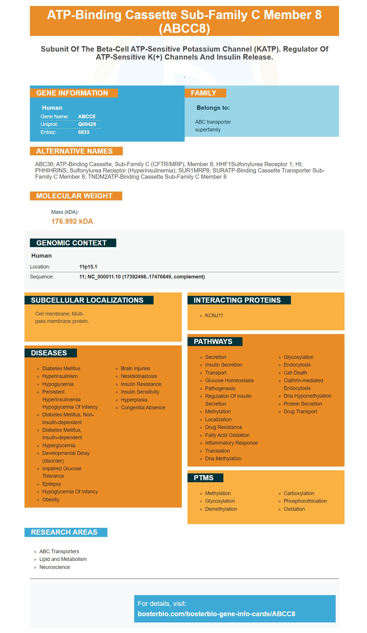


Facts about ATP-binding cassette sub-family C member 8.

.
| Human | |
|---|---|
| Gene Name: | ABCC8 |
| Uniprot: | Q09428 |
| Entrez: | 6833 |

| Belongs to: |
|---|
| ABC transporter superfamily |

ABC36; ATP-binding cassette, sub-family C (CFTR/MRP), member 8; HHF1Sulfonylurea receptor 1; HI; PHHIHRINS; sulfonylurea receptor (hyperinsulinemia); SUR1MRP8; SURATP-binding cassette transporter sub-family C member 8; TNDM2ATP-binding cassette sub-family C member 8
Mass (kDA):
176.992 kDA

| Human | |
|---|---|
| Location: | 11p15.1 |
| Sequence: | 11; NC_000011.10 (17392498..17476849, complement) |
Cell membrane; Multi-pass membrane protein.


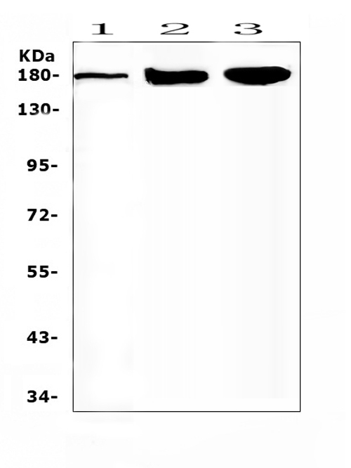
The ABCC8 Marker is one the most popular antibodies that Boster provides. You can use it to test many different samples and applications. Here are some of the benefits of using this marker. For more information, visit Boster's bio page. Find out more about Boster and its products, as well as gene infographics. You can also submit your results to receive credits for Boster's products. This program is open to scientists across the globe and not restricted to a specific country.
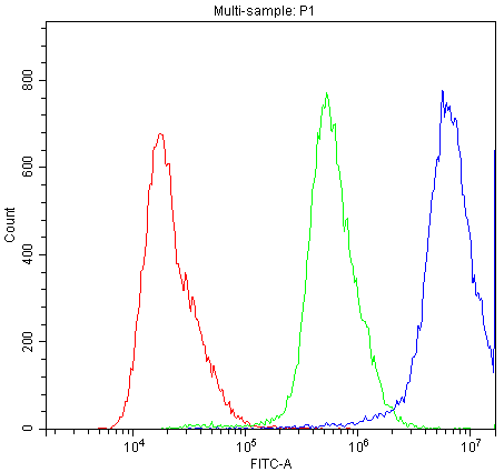
Boster's primary antibodies must be colocalized with GFP when employed in fluorescent immunocytochemistry. Colocalization refers to both labels being present at the same location, and not binding to the same protein. This is important for the use of fluorescent immunocytochemistry, since the resolution of a microscope isn't enough to determine the exact location of one specific protein. Here are some tips to make use of Boster primary antibody in fluorescent immunocytochemistry.
In order to check whether an antibody is specific to an antigen, it must be able to bind to an epitope of the antigen. Utilizing a genetic approach, a knockout animal is used to manipulate antigen expression. The tissue of knockout animals can be fixed and prepared in the same manner as experimental samples. This marker is constructed from an antigen whose expression was blocked by nonfunctional genes.
Fluorescent mIHC has numerous advantages however it is limited to two markers due to filter sets. Fluorescent mIHC uses three primary antibodies as well as fluorescent secondary antibodies to increase amplification. Primary antibodies that are raised in various species, concentrations, and isotypes are used. mIHC is a powerful tool for immunohistochemistry. While traditional mIHC relies on antibodies produced by different species, fluorescent mIHC utilizes multiple markers and requires primary antibodies of various isotypes.
Secondary antibodies used in a primary-secondary system are often double-labeled. This dual-labeling method allows for more questions to be asked of every specimen, as well as more contextual information to back up the answers. The dual-labeling system also permits for a variety in the uses for the primary antibodies. Researchers can make use of one antibody to identify various types of proteins or cells. By using two primary antibodies, a greater range of questions can be addressed and the results will be more reliable.
Since the primary antibodies are cross-conjugated it's hard to distinguish between different species with just one antibody. However, secondary antibodies utilize the ABCC8 marker, which makes the detection much easier. High-affinity secondary antibodies are great for detecting abundant protein targets and compensating for low levels. It is also compatible with other species as well as host species. It's a great solution to stain multicolor.
The ABCC8 Marker is a useful instrument to aid in cancer research and diagnostics. It can help doctors identify those suffering from tumors that are located in their brains. The levels of its expression in patients with gliomas or other cancers are greater in these two types of tumors than patients who are not affected by it. It may also assist researchers in understanding the molecular factors that are responsible for the development of these types of tumors. In one study, the ABCC8 expression levels in gliomas was compared by using the Chi square test. The results indicated that high levels of expression were associated with low WHO grade and molecular grade as well as a 1p19q combination deletion.
PMID: 21671119 by Schmid D., et al. An abundant, truncated human sulfonylurea receptor 1 splice variant has prodiabetic properties and impairs sulfonylurea action.
PMID: 7716548 by Thomas P.M., et al. Mutations in the sulfonylurea receptor gene in familial persistent hyperinsulinemic hypoglycemia of infancy.