This website uses cookies to ensure you get the best experience on our website.
- Table of Contents
Flow cytometry and Fluorescence-Activated Cell Sorting (FACS) are indispensable tools in biomedical research and clinical diagnostics. Despite their widespread use, confusion often arises regarding their terminology and functionalities. In this article, we identify distinctions between flow cytometry and FACS, and discuss their principles and applications.
Developed in the 1950s and 1960s, flow cytometry revolutionized cell analysis by allowing rapid, high-throughput measurement of multiple cellular characteristics. This technique analyzes the physical and chemical characteristics of particles or cells in a fluid suspension, and involves passing a cell-containing fluid stream through a laser beam, measuring the scattered and fluorescent light emitted by the cells.
Key aspects of flow cytometry include:

Immunohistochemistry (IHC) is a vital technique in biomedical research and clinical diagnostics, enabling the visualization and localization of specific proteins within tissue samples. In this blog, we outline the different types of IHC staining, including direct and indirect approaches, immunofluorescence, and chromogenic techniques. We also discuss the general process of IHC staining. This overview serves as an introductory guide for understanding and implementing IHC protocols.
Immunohistochemistry (IHC) staining is broadly categorized into several types based on the staining mechanism and the visualization methods used.
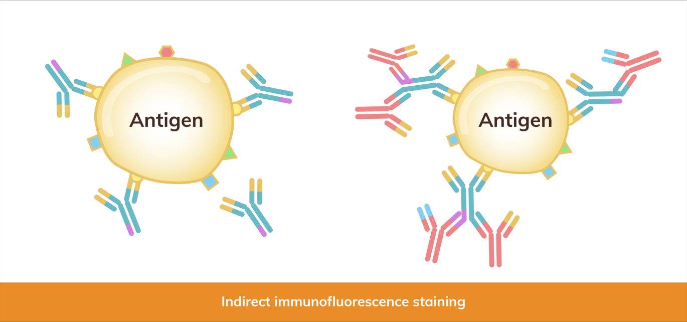
Direct Immunohistochemistry (Direct IHC): Involves the direct conjugation of the primary antibody with a detectable label (e.g., enzyme, fluorophore). This method is straightforward but may result in lower sensitivity compared to indirect methods.
Indirect Immunohistochemistry (Indirect IHC): Uses a secondary...
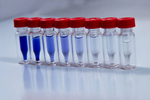
Antibodies, also known as immunoglobulins, are specialized proteins produced by the immune system to identify and neutralize foreign invaders such as bacteria, viruses, and toxins. Their ability to recognize and bind to specific antigens make them play a critical role in the immune response and serve as key tools in biomedical research.
The intricate structure of antibodies allows them to perform with high specificity and efficiency. Central to this structure are the heavy and light chains...
Immunoglobulins, commonly known as antibodies, play a pivotal role in the immune system's defense against pathogens. These specialized glycoproteins are produced by B cells and are essential for recognizing and neutralizing foreign substances, such as bacteria and viruses, through their antigen-binding sites.
In biomedical research, immunoglobulins are indispensable tools with their ability to bind specifically to antigens. Antibodies are utilized to detect and quantify proteins, pathogens...
The 2024 AACR Annual Meeting is coming up soon! Join us in San Diego from April 5-10 for the annual event. Every year, the cancer research community gathers to share the latest advances in cancer science and medicine at the AACR Annual Meeting.
Drop by Booth #804 to pick up technical resources and an adorable Dr. Booster figurine! Come and discover how our products and services can boost your research. We look forward to seeing you there...

The immune system is a complex network that protects the body from harmful pathogens such as bacteria, viruses, and other foreign substances. Central to this defense mechanism are various molecular components, including antibodies, antigens, haptens, epitopes, and immunogens. Each of these components plays a unique role in the immune response, and understanding their differences is crucial for comprehending how the immune system functions.
Antibodies, also known as immunoglobulins, are specialized Y-shaped proteins produced by B cells, a type of white blood cell. These proteins are critical in the immune response as they identify and neutralize foreign objects like bacteria and viruses. The structure of antibodies allows them to specifically recognize and bind to antigens, effectively marking them for destruction by other immune cells. This specificity is due to the unique binding sites on the antibody that match specific parts of the antigen, known as epitopes.
Antigens are molecules or molecular structures, often found on the surface of pathogens, that can elicit an immune response. They are typically proteins or polysaccharides and are recognized by the immune system as foreign. The interaction between antigens and antibodies is highly specific, akin to a lock and key, where the antibody binds to the antigen to neutralize or flag it for removal. This interaction is fundamental to the body’s ability to recognize and remember pathogens, forming the basis of immunity.
A fascinating aspect of immune recognition involves haptens. Haptens are small molecules that, by themselves, are not immunogenic, meaning they cannot independently provoke an immune response. However, when haptens bind to larger carrier proteins, they form a complex that the immune system recognizes as foreign, triggering an immune response. This phenomenon is particularly important in pharmacology and allergen studies, where certain drugs or small molecules can act as haptens, causing allergic reactions when they bind to proteins in the body.
Epitopes, also known as antigenic determinants, are specific regions on an antigen that are recognized by antibodies. Each antigen can have multiple epitopes, allowing for the binding of different antibodies. This multiplicity enables a robust and versatile immune response, as various antibodies can target different parts of a single pathogen. The specificity of epitopes is crucial in vaccine development, as vaccines aim to introduce harmless forms of epitopes to stimulate an immune response without causing disease.
Immunogens are a subset of antigens that can trigger an immune response on their own. While all immunogens are antigens, not all antigens qualify as immunogens. For instance, a hapten is an antigen but not an immunogen unless it is attached to a carrier protein. Immunogens must have certain properties, such as adequate size and molecular complexity, to effectively stimulate the immune system. This distinction is important in vaccine development and allergy research, where understanding what makes an antigen immunogenic can inform treatment strategies.
We’ve provided a table below that summarizes and highlights the distinct roles and interactions of these immune system components.
| Term | Description | Example |
|---|---|---|
| Antibody | Y-shaped proteins produced by B cells that specifically recognize and bind to antigens, aiding in the neutralization and destruction of pathogens. | IgG, IgM, IgA |
| Antigen | Substances, often proteins or polysaccharides on the surface of pathogens, that trigger the production of antibodies and stimulate an immune response. | Viral proteins, bacterial cell wall components |
| Hapten | Small molecules that are not immunogenic on their own but can elicit an immune response when bound to a larger carrier protein. | Penicillin, certain drugs |
| Epitope | Specific regions on antigens that are recognized and bound by antibodies. | Binding site on a viral protein |
| Immunogen | A type of antigen capable of independently provoking an immune response, possessing the necessary properties to stimulate immune cells. | Whole viruses, bacterial toxins |
The immune system's effectiveness hinges on interactions among antibodies, antigens, haptens, epitopes, and immunogens. Antibodies recognize and bind to antigens, identifying foreign molecules for destruction. Haptens, though non-immunogenic alone, provoke responses when linked to larger proteins. Epitopes are specific antigen regions targeted by antibodies, while immunogens independently trigger immune responses. Comprehending these dynamics not only advances immunological understanding but als...
Meet us in Salt Lake City from March 10-14 at the Society of Toxicology (SOT) 63rd Annual Meeting and ToxExpo! Over 5,000 toxicologists and professionals attend this event to share the latest science and technology advancements, make new connections, and engage in professional development.
Stop by Booth #1802 to pick up technical resources and an adorable Dr. Booster figurine! Come and discover how our products, services, and resources can boost your research. We hope to see you there...

Blotting techniques are essential tools in molecular biology and biochemistry, allowing researchers to detect and analyze nucleic acids and proteins. These techniques enable scientists to uncover intricate details about genetic material, gene expression, and protein interactions. From the foundational Southern blotting, which revolutionized DNA analysis, to the versatile Western blotting, a staple in protein research, blotting methods have become indispensable tools in the laboratory.
This blog will delve into 10 blotting types, each with unique applications and methodologies. We'll explore the nuances of each technique, highlighting their roles in studying DNA, RNA, and proteins.
If you’re looking to learn about the different types of blotting techniques for your research, this guide is for you!
Southern blotting, the original blotting technique, is named after its inventor Edwin Southern who developed the method in 1975 for transferring DNA from a gel to a membrane, enabling the identification of specific DNA sequences. The naming convention for subsequent blotting techniques was influenced by this original name.
To this day, Southern blotting remains a common technique for DNA analysis and identification. The process involves digesting DNA with restriction enzymes, separating the fragments by gel electrophoresis, and transferring them onto a membrane. Labeled DNA or RNA probes then hybridize to the target sequences. Popular in genetics and molecular biology, Southern blotting is used for the detection of specific DNA sequences, study of DNA methylation patterns, RFLP (Restriction Fragment Length Polymorphism) analysis for genetic fingerprinting, identification of gene mutations and polymorphisms, and gene mapping and cloning.
Northern blotting was named by James Alwine, David Kemp, and George Stark in a 1977 paper as a playful pun on Southern blotting, indicating its use for RNA rather than DNA.
This technique is similar to Southern blotting, but focuses on RNA analysis. RNA samples are separated by gel electrophoresis, transferred to a membrane, and hybridized with labeled DNA or RNA probes to detect and quantify specific RNA transcripts. Northern blotting is commonly used to analyze gene expression patterns, determine mRNA size and abundance, study RNA processing and degradation, and detect alternative splicing events.
Western blotting, also called immunoblotting, is widely used for protein detection and analysis, making it the most popular blotting technique. The method was first described by Towbin et al. in 1979 and the term "western" was coined by W. Neal Burnette in 1981 as a tongue-in-cheek reference to the direction naming theme established by Southern and Northern blotting, this time focusing on proteins instead of nucleic acids.
This technique detects specific proteins by separating them via gel electrophoresis (typically SDS-PAGE), transferring them to a membrane (usually PVDF or nitrocellulose: AR0135-02, AR0135-04), and using primary and secondary antibodies for detection. The secondary antibody is usually conjugated to an enzyme or a fluorescent tag that produces a detectable signal when exposed to a substrate or under specific conditions. For your western blot experiment, you can explore Boster’s catalog to browse primary antibodies and secondary antibodies validated for western blot as well as find western blot reagents you will need.
In protein research and diagnostics, Western blot is used for the detection and quantification of specific proteins in a sample, analysis of protein expression levels across different conditions or treatments, detection of post-translational modifications (e.g., phosphorylation, glycosylation), study of protein-protein interactions, and confirmation of protein identity and purity in recombinant protein production.
To learn more about western blotting, download our Western Blot eBook, which discusses the principle, protocol, troubleshooting tips, and FAQs for western blot.
Following the directional theme set by Southern and Western blotting, Eastern blotting is used to analyze post-translational modifications (PTMs) of proteins, such as glycosylation or phosphorylation. Many scientists deem Eastern blotting as a variation of Western blotting.
The Eastern blot technique involves transferring proteins separated by gel electrophoresis onto a membrane, followed by detection using specialized probes or antibodies targeting the PTM of interest. Though less frequently performed than other blotting techniques, Eastern blotting is useful for analyzing PTMs (e.g., glycosylation), studying protein modifications (e.g., phosphorylation, lipidation), and detecting and characterizing glycoproteins and other modified proteins.
Far-Western blotting, derived from Western blotting, focuses on protein-protein interactions. It involves transferring proteins separated by gel electrophoresis onto a membrane and identifies interactions using labeled proteins or peptides to probe for binding with the immobilized target proteins.
Growing in popularity, Far-Western blotting is valuable for identifying and studying protein-protein interactions, mapping interaction domains, and screening potential binding partners.
Southwestern blotting combines features of Southern and Western blotting techniques, focusing on DNA-binding protein detection. This technique involves transferring denatured proteins from a gel onto a membrane, followed by incubation with labeled DNA probes to identify proteins that bind to specific DNA sequences.
Though less common than Southern and Western blotting, Southwestern blotting is useful for detecting DNA-binding proteins, studying protein-DNA interactions, and identifying transcription factors and other regulatory proteins.
Reverse Northern blotting indicates the reversal of the Northern blotting process. Instead of transferring RNA and probing with DNA, Reverse Northern blotting transfers DNA and probes with labeled RNA.
This technique involves immobilizing DNA on a membrane and hybridizing it with labeled RNA or cDNA probes to detect DNA sequences. Although less common than standard Northern blotting, it is used for gene expression analysis using cDNA or genomic DNA arrays, screening differentially expressed genes in various conditions, and studying changes in transcript levels in response to treatments.
Colony blotting is named for its application in screening microbial colonies, such as bacterial or yeast colonies. It involves transferring entire colonies from a culture plate onto a membrane, where specific nucleic acids or proteins are detected through DNA hybridization analysis.
Primarily used in microbiology and cloning, colony blotting helps screen colonies for target DNA or RNA sequences, identify recombinant clones containing specific genetic inserts, and rapidly detect plasmid-containing colonies.
Dot blotting is frequently used as a quick and simple screening method for the presence or absence of nucleic acids and proteins. Named for the dot-like application of samples on the membrane, it is a simplified version of Western blotting, where samples are directly spotted onto a membrane for detection without prior gel electrophoresis.
Popular for rapid screening, dot blotting is used to screen specific nucleic acids or proteins, relatively quantify target molecules, and analyze large numbers of samples simultaneously. It should be noted that dot blots do not provide information about molecular weight, so false positive signals or the presence of modified proteins are difficult to identify.
Slot blotting is similar to dot blotting, but less commonly used. Named for the slot-like application of samples, slot blotting involves applying samples in rectangular slots on a membrane, allowing more uniform application and the quantification of target molecules in a sample without the need for gel electrophoresis.
This technique is helpful for analyzing nucleic acids or proteins without electrophoresis, comparing the relative abundance of target molecules in different samples, and screening multiple samples in high-throughput formats. As with dot blots, slot blots are also unable to provide information about the size of the target protein.
We have provided a table below that highlights the features of each blotting type.
| Blotting Type | Target Molecule | Detection Method | Technique Description | Applications |
|---|---|---|---|---|
| Southern Blotting | DNA | Labeled DNA/RNA probes | DNA fragments are separated by electrophoresis, transferred to a membrane, and probed. | Detection of DNA sequences, genetic fingerprinting, gene mapping |
| Northern Blotting | RNA | Labeled DNA/RNA probes | RNA is separated by electrophoresis, transferred to a membrane, and hybridized with probes. | Analysis of gene expression, RNA processing, alternative splicing |
| Western Blotting | Proteins | Primary/secondary antibodies | Proteins are separated by SDS-PAGE, transferred to a membrane, and detected with antibodies. | Protein detection, expression analysis, post-translational modifications |
| Eastern Blotting | Modified proteins | Specific probes/antibodies | Proteins are transferred to a membrane and probed for post-translational modifications. | Analysis of glycosylation and other post-translational modifications |
| Far-Western Blotting | Proteins (interactions) | Labeled proteins/peptides | Proteins are transferred to a membrane, probed with labeled proteins to detect interactions. | Study of protein-protein interactions, mapping interaction domains |
| Southwestern Blotting | DNA-binding proteins | Labeled DNA probes | Proteins are transferred to a membrane and probed with labeled DNA to detect binding. | Detection of DNA-binding proteins, study of protein-DNA interactions |
| Reverse Northern Blotting | DNA | Labeled RNA/cDNA probes | DNA is immobilized on a membrane and hybridized with labeled RNA/cDNA probes. | Gene expression profiling, screening for differentially expressed genes |
| Colony Blotting | DNA/RNA in colonies | Labeled probes | Microbial colonies are transferred to a membrane and probed for specific sequences. | Screening for specific sequences, identifying recombinant clones |
| Dot Blotting | Nucleic acids/proteins | Labeled probes/antibodies | Samples are spotted directly onto a membrane for rapid detection. | Rapid screening, quantification of target molecules |
| Slot Blotting | Nucleic acids/proteins | Labeled probes/antibodies | Samples are applied in slots on a membrane for quantitative analysis. | Quantitative analysis, comparing relative abundance of target molecules |
Blotting techniques are important tools in molecular biology and biochemistry. Each blotting method caters to different experimental needs, enabling detailed analysis of DNA, RNA, proteins, and their interactions. By utilizing each technique’s unique advantages and applications, researchers can deepen our understanding of molecular interactions and processes.