This website uses cookies to ensure you get the best experience on our website.
- Table of Contents
As of January 2022, there are nearly 2,500 brain-derived neurotrophic factor (BDNF) antibodies to choose from according to CiteAb. In this article, I will share how I found the BDNF antibody that worked for my projects and I hope my experience is helpful if you are currently searching for good BDNF antibodies. I will also discuss the expected BDNF molecular weight in Western blots, BDNF positive and negative control designs, and a step-by-step guide of how I currently search for antibodies.
During my first year in the lab, my team was investigating BDNF’s role in Rett syndrome by treating various CNS cells with inflammatory cytokines in vitro and we discovered upregulation of BDNF mRNAs. Since we had to conduct protein level analysis, I started the hunt for anti-BDNF antibodies.
Almost any scientist who has worked with antibodies knows that many unexpected issues can occur even for the most well-known biomarkers and established antibodies. To prepare for the antibody search, I generally get 2 facts sorted out first:
These facts will come in handy later to help rule out antibodies (in my case, anti-BDNF antibodies) that have incorrect WB band sizes and poorly designed controls.
(If you are already familiar with BDNF, you can skip this section) BDNF stands for brain-derived neurotrophic factor, which is involved in modulating activities in the central nervous system and peripheral nervous system. The protein has 5 isoforms and 1 cleavage site, which determines its molecular weight in Western blotting. The uncleaved BDNF is called pro-BDNF (precursor protein), which can be cleaved into a pro-peptide and mature BDNF (about half the size of pro-BDNF). Later in this article, I will discuss BDNF expressing tissues and cells. You can also check out this short 3 min video covering the essential facts about BDNF.
I typically follow similar steps described in this guide when searching for antibodies. First, figure out all the synonyms/aliases of the biomarker. Then, much like selecting job candidates, start screening with some criteria and filters based on your project’s needs. In most cases, however, you do not have the time to investigate every antibody. For example for BDNF alone, there are 2000+ antibodies from 50+ companies.
Marketplaces like Biocompare, Antibodypedia, CiteAb, Linscott’s Directory, Antibody Resources, and Labome are great starting points. Platforms like GeneCards are also helpful, but it lacks the filter search function and requires you to manually check all options.
For your protein of interest, you should know its expected molecular weight in Western blot, and the expected tissue type and subcellular localization for immunohistochemistry (IHC), which will help you tell the good from the bad when you review details for each commercially available antibody. Uniprot, NCBI Entrez, and Human Protein Atlas are 3 great databases for this purpose.
Lastly, research which tissues and cell types to use for positive and negative controls before beginning your antibody search. Ideally, you want protein level evidence, but often it is unavailable. In that case, you may have to settle for mRNA level evidence. Uniprot.org and Proteinatlas.org are my favorite sites for researching such information.
Note: If the antibody is only validated on purified recombinant protein/peptide, it means little since there is low correlation between how an antibody reacts with a recombinant protein and how it reacts with biological samples.
It is better to know the expected molecular weight of BDNF for Western blot before reviewing various antibodies. BDNF can be cleaved into two chains which could lead to multiple bands on a Western blot, as well as false high readings in ELISA. BDNF is a single chain protein 247 AAs long (precursor), including signal peptide. It is worth noting that BDNF is cleaved by MBTPS1 at AA 57-58.1 This turns BDNF from its precursor form into its mature form, which is 119 AAs long. It is important to clarify whether your antibody or ELISA kit is specific to the mature form or can recognize both the precursor and the mature BDNF. If you see two bands in your WB, one around 30 kDa and one at around 14 kDa, that’s pro and mature forms of BDNF at work.
Isoforms are a common cause of multiple bands in Western blotting, but not in the case of BDNF. There are 5 isoforms for BDNF, and all are of similar sizes. After reviewing BDNF Western blot images online, it appeared that these isoforms do not seem to cause much problem in terms of extra bands in the blots. My educated guess is that these isoforms exist at much lower levels in the sample matrix, though I have no direct evidence for this claim.
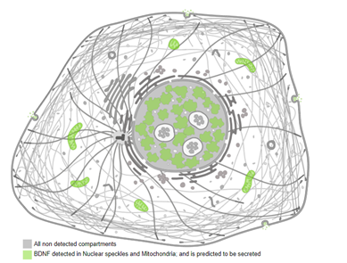
Since BDNF is secreted, mRNA levels are poor indicators of where the protein might be present. Non-BDNF-producing cells and body fluids could still have high levels of BDNF secreted by other cells. According to Uniprot, BDNF is detected in blood plasma and saliva. It is expressed in the central nervous system (CNS), mostly by hippocampus, amygdala, cerebral cortex, and cerebellum. It is also expressed at lower levels in the heart, lungs, skeletal muscles, testes, prostate, and placenta. (BDNF: Uniprot, Protein Atlas). Since it is a secreted protein, subcellular staining can be expected in cell membranes. It is also concentrated in mitochondria and nuclear speckles.
There are several tissues and cell types known to highly express BDNF, so positive control design should be simple. As for negative control, there is not a set of widely used negative tissues/cell types in publications. I did find a few cell lines cited in publications. There was also mRNA evidence for BDNF negative cell lines on Protein Atlas, as shown in the image below:
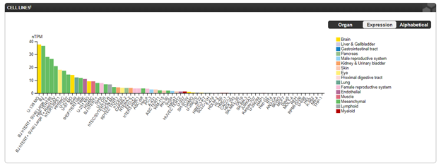
In a publication, A-431 was used as BDNF negative control, as per antibody manufacturer’s suggestion, and it worked well even though A-431 is shown to have a low level of BDNF mRNA expression in the image above.2 This suggests that any cell lines to the right of A-431 in the image above have the potential to be a good negative control for BDNF in Western blotting. Some validation will be needed to substantiate cell lines that have not been used before. Since A-431 has been validated as a negative control, I would recommend it. When checking validation images by vendors, you should reference the tissues and cell lines they used in Western blotting to see if they match up with mRNA levels. Though the correlation is not 100%, too much deviation would raise a red flag.
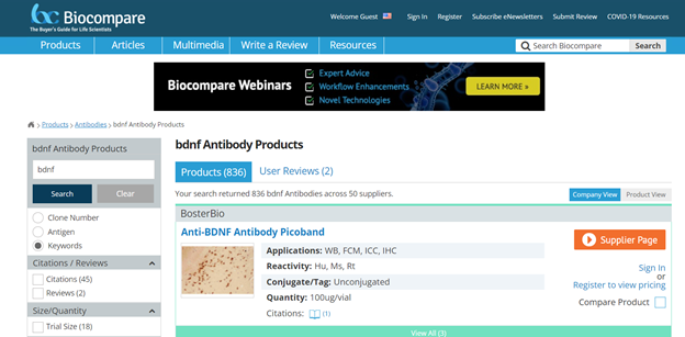
Since I knew the expected BDNF band size in Western blot, and which tissues and cells positively and negatively express BDNF, I began the hunt. A Biocompare search for BDNF antibodies revealed 836 antibodies from over 50 companies.
Next, I used filters to narrow down the choices. Ideally, I wanted no more than 10-20 antibodies to manually review. In my case, I needed the antibody to work both in Western blotting and IHC while being reactive against mouse. I also checked “Citations” as a filtering factor. With all these conditions, there were only 28 candidates left.
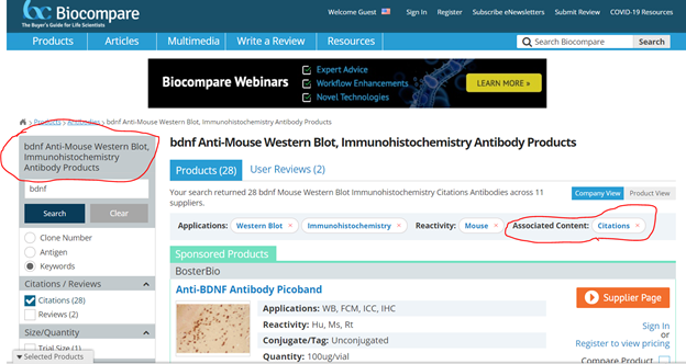
I could have further narrowed it down by checking “Flow Cytometry” under “Applications”; however, most antibody companies do not routinely validate with Flow cytometry, so I chose to review this aspect manually as it was not a deal breaker if the antibody is not validated on Flow.
It is worth noting that the default filter logic on Biocompare for within the same attribute is the “OR” logic. This means that if you select western blot and immunohistochemistry, it will show you antibodies that have either WB or IHC. If you further select additional applications, it will broaden search results, which may not be what you want, so please be mindful when choosing filters.
Even though anecdotal evidence is not ideal for decision making, there is always a place for it. If you are somewhat new to antibody hunting, it would certainly help to ask your labmates and colleagues about what experience they have had with any of the companies you consider to purchase from. Be ready to tune out weak indicators, such as, “I tried xx (company) antibody once, but the results were only OK.” Look for strong indicators, such as, “The customer service from XXX is amazing/terrible” or “I have used a few antibodies from XXX and they were all good/bad”.
To avoid bias, I am going to skip sharing the anecdotes I received. However, my peers’ recommendations were certainly helpful in further narrowing down the antibody candidate pool and avoiding some companies I wanted to purchase from.
Now comes the fun (or tedious) part. I had narrowed down the antibody candidates as much as I could, and I was ready to look at the fine print. Generally, I primarily consider 3 things:
I look at the manufacturer-provided Western blot images’ band sizes to see if they match up with expectations. I also look at the IHC images and check if the signal-to-noise ratio is high enough and whether the staining pattern is as expected. If the manufacturer does not have a Western blot image tested with real tissues/cells (e.g. supplier has only tested antibody on recombinant proteins), it is a hard pass. If there are no better options available and you are desperate to get an antibody, be sure to contact the company and secure a refund guarantee or a small sample size before ordering.
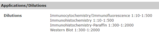
I look at the manufacturer’s suggested dilution ratios and what dilution ratios they used for their own images. If the dilution ratio is too low, it may indicate that the antibody’s affinity towards the intended antigen is low. Non-specific binding is common and occurs at a similar strength across different clones. Having a low specific-affinity means the biomarker might not be detected in the samples with low concentrations and there could be a higher chance for non-specific binding. Typically, a working concentration around 1ug/ml is considered high affinity. If it takes 5ug/ml or higher concentration to work, the chances of non-specific binding increase significantly. Sometimes, a company might not disclose the antibody concentration. You can contact them to find out or make an educated guess. In my experience, the most common antibody concentrations fall between 0.3 mg/ml to 2 mg/ml. For example, a 1:300 dilution ratio may indicate that the antibody does not have high affinity, or the company put very little antibody in the vial. However, 1:1000 to 1:5000 dilution ratios could be good signs. These numbers are just recommendations because concentration may vary greatly due to several factors, such as the format of the antibodies or the lab protocol. The goal is to be mindful of the antibody concentration and avoid low affinity antibodies.
If there are still enough candidates left, I would look through the publications citing these antibodies, and review the published IHC images for staining patterns as well as the Western blot images to check if the antibodies consistently generated clear bands at the right molecular weights across different biological matrices.
After reviewing several BDNF Western blots (images below), here are my conclusions: BDNF has a pro-form (~30 kDa) and a mature form (~14 kDa). The BDNF precursor is expressed in many cells, mostly in the CNS but also in places like the liver (For more detailed information, see section “BDNF positive control and negative control design”). In the cells/tissues known to express BDNF, we need to understand how much precursor will be present in relation to mature BDNF. When proBDNF and mature BDNF both exist in those particular cells/tissues, we will observe 2 BDNF bands with similar magnitude. However, in tissues and cells that do not produce BDNF but receive BDNF from the endocrine system, such as platelets, there should only be mature BDNF present; thus, a single band near 14 kDa. Below, I review some of the BDNF antibodies I was considering to use for my research.
The Abcam BDNF antibody (ab108319) Western blot image shows multiple bands. The band near 30 kDa can be explained as precursor, but a few bands are likely non-specific.
The official description from Abcam’s BDNF antibody is
“Predicted band size: 15 kDa, Additional bands at: 15 kDa (possible mature (processed) protein), 28 kDa (possible multimer), 35 kDa, 45 kDa (possible immature (unprocessed)). We are unsure as to the identity of these extra bands.”
--Source: abcam.com
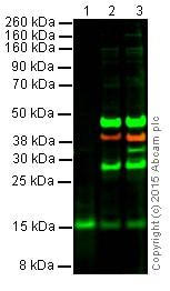
Most dimerizations do not survive the harsh conditions of Western blotting. While plausible, I have not found much documentation of BDNF dimers or multimers being observed in WB. Between non-specific binding and multimer theory, the former seems more likely. Of course, we are still in the antibody research phase, so all these deductions are only educated guesses, and none are experimentally proven.
The Novus Bio BDNF antibody (NB100-98682SS) Western blot image shows single bands at 14 kDa. Since the sample type is human platelet, which is a receiver of BDNF and not an expressor, the band size is reasonable. The dilution ratio for the primary antibody is 1:1000, which is not bad. What’s really nice is the CYP-induced cystitis validation the company performed. According to Novus Bio, cystitis is expected to increase the amount of BDNF in platelets, and the image has shown so accordingly. I am not that familiar with cystitis and how its inflammatory characteristics affect BDNF, but this image seems plausible. I assumed someone had put in a lot of thought into designing this validation and accepted its conclusion. If you are an expert on cystitis and notice anything worth mentioning, please feel free to leave a comment.
One imperfection is the dilution ratios of Novus Bio BDNF antibody, which could be better. The suggested ratios are broad, ranging from 1:10 to 1:500. If they could be a little more specific, given they have validated with mouse hippocampus, that would have been helpful. The wider the range, the more optimization one has to do before one can use the antibody. The lowest suggested dilution ratio is only 1:10 for IHC. When high levels of antibodies are added to IHC and immunofluorescence, the likelihood of non-specific binding can also be high.
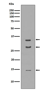
The Boster Bio BDNF antibody’s Western blot image shows 3 bands, 2 at the expected locations near 30kDa and 14 kDa. There is another band at around 34 kDa, which could be a non-specific band.
It is worth noting this is a rabbit monoclonal BDNF antibody. Rabbit monoclonal antibodies are known to have high specificity, and have a reputation of working well for IHC and IF staining.
 |
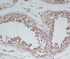 |
As shown in this image, the IHC image looks quite decent, showing signal specifically in the target cells (cytoplasm and membrane) with a clean background.
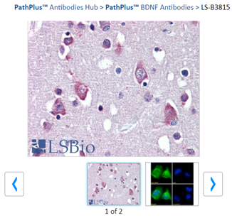
At first glance, I noticed this antibody has no Western blot image. Since we have a few great alternatives to choose from, we should stay with those that have proper evidence for the antibody’s specificity.
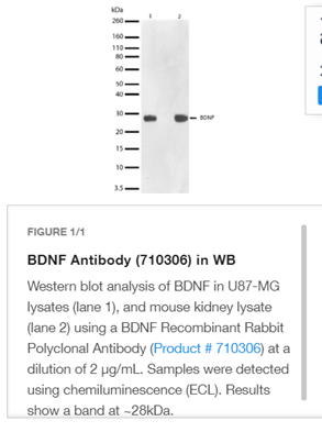
The only image for this antibody is a Western blot showing a band at 30 kDa, but it is missing the 14 kDa band. I did not find evidence that the cell line U87-MG or mouse kidney does not express mature BDNF. However, on this antibody’s page, it states that the antigen is “Recombinant protein corresponding to amino acids 129–247 of human BDNF”, which is the region for mature BDNF. Thus, it is likely this antibody is unable to detect mature BDNF as it was intended.
After some efforts, I was able to narrow down the BDNF antibody candidates to 2 companies, Novus Bio and Boster Bio. Novus Bio exceled at validating the antibody using CYP-treated cells. Boster Bio has a rabbit monoclonal with a clear IHC image. I am sure there are more BDNF antibodies on the market that are of good quality as well. If you have a good BDNF antibody to recommend, feel free to leave a comment.
Empower your research and extract crucial insights from your samples with our exceptional service. Don't miss out—contact us now to discuss your project requirements and see how our premium Western blotting service can revolutionize your work.