This website uses cookies to ensure you get the best experience on our website.
- Table of Contents

Understanding the relative abundance of target proteins and effectively normalizing data are crucial aspects of Western blot analysis. While loading control antibodies have long been the gold standard, recent advancements have highlighted the potential advantages of using total protein stain (TPS) as an alternative approach.
Boster Bio provides...
Western blotting (also called Protein Immunoblotting) is an analytical technique used to detect specific proteins in the given sample. It uses SDS-polyacrylamide gel electrophoresis (SDS-PAGE) to separate various proteins contained in the sample. The separated proteins are then transferred or blotted onto a matrix, where they are stained with antibodies specific to the target protein. Expression details of the target proteins in the given cells or tissue homogenate can then be obtained through analyzing the location and intensity of the specific reaction. Western blotting analysis can detect target protein as low as 1 ng due to high resolution of the gel electrophoresis and strong specificity and high sensitivity of the immunoassay. This method is used in the fields of molecular biology, biochemistry, immunogenetics and other molecular biology disciplines for various experiments.
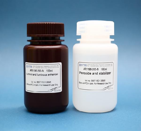
Enhanced Chemiluminescence (ECL) Western Blot Substrate is a very sensitive, non-radioactive, enhanced luminol-based chemiluminescent substrate that allows for easy detection of horseradish peroxidase (HRP) on immunoblots. HRP is a common molecule conjugated to antibodies. ECL Western Blot Substrate has the capability of offering...
Western blotting (also called Protein Immunoblotting) is an analytical technique used to detect specific proteins in the given sample. It uses SDS-polyacrylamide gel electrophoresis (SDS-PAGE) to separate various proteins contained in the sample. The separated proteins are then transferred or blotted onto a matrix, where they are stained with antibodies specific to the target protein. Expression details of the target proteins in the given cells or tissue homogenate can then be obtained through analyzing the location and intensity of the specific reaction. Western blotting analysis can detect target protein as low as 1 ng due to high resolution of the gel electrophoresis and strong specificity and high sensitivity of the immunoassay. This method is used in the fields of molecular biology, biochemistry, immunogenetics and other molecular biology disciplines for various experiments.
Filter paper (or blotting paper) is important to ensure quick and efficient transfer of molecules from the electrophoresis gel to the matrix membrane. It is used for transfer sandwiches and cassettes during Western blotting and assists with transferring proteins unto polyvinylidene fluoride (PVDF), nitrocellulose, and other types of membranes. Western blot filter paper is a semi-permeable paper barrier used to separate fine solid particles from liquids...

Nitrocellulose membranes are one of the top matrices used in protein blotting. They have high protein-binding affinity, compatibility with a variety of detection methods, and the ability to immobilize proteins, glycoproteins, or nucleic acids. Examples of compatible detection methods include chemiluminescence, chromogenic, and fluorescence. It is proven to produce excellent signal-to-noise results when used for amino acid analysis and western, northern, and Southern blotting.
Western blotting (also called Protein Immunoblotting) is an analytical technique used to detect specific proteins in the given sample. It uses SDS-polyacrylamide gel electrophoresis (SDS-PAGE) to separate various proteins contained in the sample. The separated proteins are then transferred or blotted onto a matrix, where they are stained with antibodies specific to the target protein. Expression details of the target proteins in the given cells or tissue homogenate...

In order to get the best results from your ELISA assay, the dilution factors of the sample and the detection antibodies must be optimized. If your sample or antibodies are too concentrated, you risk saturating the assay. If they are not concentrated enough, your signal will be weak and difficult to detect. For strong, quantifiable signal, use a checkerboard titration to test for the optimal...
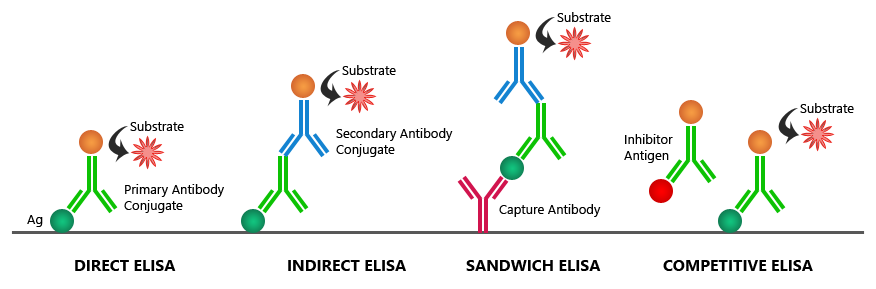
Are you familiar with the multiple methods you could use to perform an ELISA? Among the standard assay formats illustrated below, where differences in both capture and detection are in concern, it is important to differentiate between the particular strategies that exist specifically for the detection step. However an antigen is captured to the
If you are having trouble with saturated signals in your ELISA data results, check out this table for Boster’s possible solutions to your problem:
| Possible Causes | Possible Solutions |
|---|---|
| High sample concentration | Use higher sample dilutions (Determine the optimal dilutions by titration assay) |
| Excessive substrate | Decrease concentration or amount of substrate: Follow manufacturer guidelines (The substrate provided with the ELISA kit might require further dilution) |
| Substrate color changed before use | Make substrate immediately before use |
| Non-specific antibody binding | Try different formulations in coating solutions; Ensure wells are pre-processed to prevent non-specific binding; Use affinity-purified antibody and preferably one that is pre- adsorbed; Use serum (5-10%) from same species as secondary antibody (bovine serum is also recommended). |
| Incubation time too long | Follow the manufacturer guidelines (If the problem persists, try incubating samples at 4°C overnight) |
| Excess antibody | Repeat the assay with lower antibody concentrations to find the optimal one for your experiment |
| Contaminated buffers with metals or HRP | Make and use fresh buffers |
| Insufficient washing | Follow the manufacturer guidelines; At the end of ea |
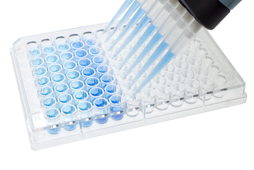
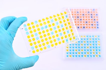
ELISA (enzyme-linked immunosorbent assay) is a plate-based assay used to detect the concentration of a specific protein in a liquid sample. Three different types of data output can be obtained:
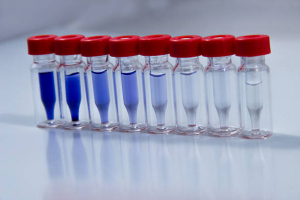
To set up a standard curve, ELISA standards should be carefully prepared for accurate sample quantification of...

ELISA (enzyme-linked immunosorbent assay) is a convenient and simple method to quantitatively or qualitatively detect peptides, proteins, antibodies, and hormones in samples, rendering it as one of the most widely used immunoassays. Despite the many advantages of conducting ELISA, there are some mistakes that could turn your ELISA experiment sour. Help prevent this situation from happening by avoiding 5 common pitfalls when performing an ELISA:
1. “I think I still have some of the TMB coloring development agent from the previous kit. Maybe I’ll use that instead.”
Stop! Wanting to conserve resources is a good habit, but not in this case. We should avoid using reagents from different batches together. Each...