Click image to see more details
Product Info Summary
| SKU: | M02659 |
|---|---|
| Size: | 100 μg/vial |
| Reactive Species: | Human, Mouse, Rat |
| Host: | Mouse |
| Application: | Flow Cytometry, IF, IHC, ICC, WB |
Customers Who Bought This Also Bought

Product info
Product Name
Anti-PDE6 beta/PDE6B Antibody Picoband™ (monoclonal, 8I2F10)
SKU/Catalog Number
M02659
Size
100 μg/vial
Form
Lyophilized
Description
Boster Bio Anti-PDE6 beta/PDE6B Antibody Picoband™ (monoclonal, 8I2F10) catalog # M02659. Tested in FCM, IF, IHC, ICC, WB applications. This antibody reacts with Human, Mouse, Rat.
Storage & Handling
At -20°C for one year from date of receipt. After reconstitution, at 4°C for one month. It can also be aliquotted and stored frozen at -20°C for six months. Avoid repeated freezing and thawing.
Cite This Product
Anti-PDE6 beta/PDE6B Antibody Picoband™ (monoclonal, 8I2F10) (Boster Biological Technology, Pleasanton CA, USA, Catalog # M02659)
Host
Mouse
Contents
Each vial contains 4 mg Trehalose, 0.9 mg NaCl and 0.2 mg Na2HPO4.
Clonality
Monoclonal
Clone Number
8I2F10
Isotype
Mouse IgG2b
Immunogen
E.coli-derived human PDE6 beta/PDE6B recombinant protein (Position: K25-Q237).
*Blocking peptide can be purchased. Costs vary based on immunogen length. Contact us for pricing.
Cross-reactivity
No cross-reactivity with other proteins.
Reactive Species
M02659 is reactive to PDE6B in Human, Mouse, Rat
Applications
M02659 is guaranteed for Flow Cytometry, IF, IHC, ICC, WB Boster Guarantee
Observed Molecular Weight
98 kDa
Calculated molecular weight
Background of PDE6 beta
Photon absorption triggers a signaling cascade in rod photoreceptors that activates cGMP phosphodiesterase (PDE), resulting in the rapid hydrolysis of cGMP, closure of cGMP-gated cation channels, and hyperpolarization of the cell. PDE is a peripheral membrane heterotrimeric enzyme made up of alpha, beta, and gamma subunits. This gene encodes the beta subunit. Mutations in this gene result in retinitis pigmentosa and autosomal dominant congenital stationary night blindness. Multiple transcript variants encoding different isoforms have been found for this gene.
Antibody Validation
Boster validates all antibodies on WB, IHC, ICC, Immunofluorescence, and ELISA with known positive control and negative samples to ensure specificity and high affinity, including thorough antibody incubations.
Assay dilution & Images
Reconstitution
Adding 0.2 ml of distilled water will yield a concentration of 500 μg/ml.
Assay Dilutions Recommendation
The recommendations below provide a starting point for assay optimization. The actual working concentration varies and should be decided by the user.
Western blot, 0.25-0.5 μg/ml, Mouse, Rat
Immunohistochemistry(Paraffin-embedded Section), 2-5 μg/ml, Rat
Immunocytochemistry/Immunofluorescence, 5 μg/ml, Human
Flow Cytometry (Fixed), 1-3 μg/1x6 cells, Human
Validation Images & Assay Conditions
Click image to see more details
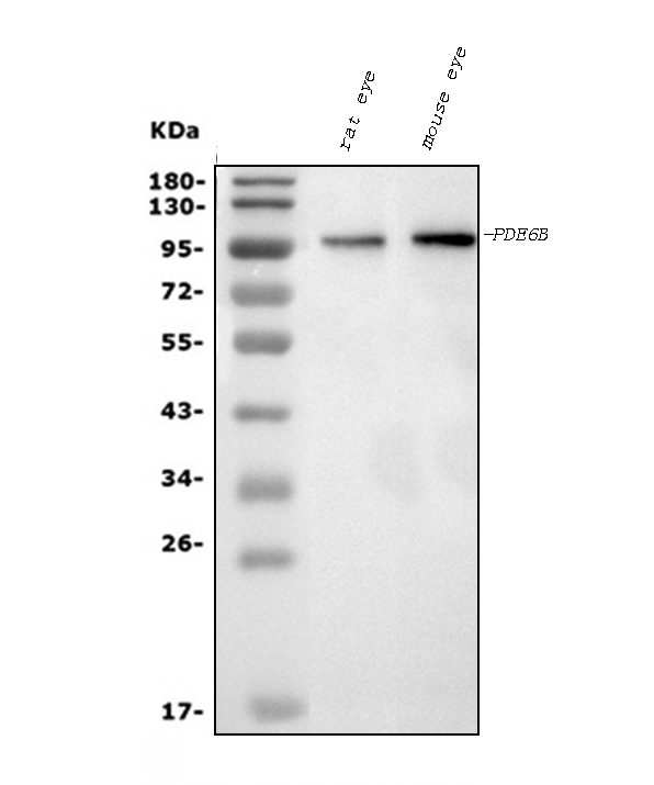
Figure 1. Western blot analysis of PDE6 beta/PDE6B using anti-PDE6 beta/PDE6B antibody (M02659).
Electrophoresis was performed on a 5-20% SDS-PAGE gel at 70V (Stacking gel) / 90V (Resolving gel) for 2-3 hours. The sample well of each lane was loaded with 30 ug of sample under reducing conditions.
Lane 1: rat eye tissue lysates,
Lane 2: mouse eye tissue lysates.
After electrophoresis, proteins were transferred to a nitrocellulose membrane at 150 mA for 50-90 minutes. Blocked the membrane with 5% non-fat milk/TBS for 1.5 hour at RT. The membrane was incubated with mouse anti-PDE6 beta/PDE6B antigen affinity purified monoclonal antibody (Catalog # M02659) at 0.5 μg/mL overnight at 4°C, then washed with TBS-0.1%Tween 3 times with 5 minutes each and probed with a goat anti-mouse IgG-HRP secondary antibody at a dilution of 1:10000 for 1.5 hour at RT. The signal is developed using an Enhanced Chemiluminescent detection (ECL) kit (Catalog # EK1001) with Tanon 5200 system. A specific band was detected for PDE6 beta/PDE6B at approximately 98 kDa. The expected band size for PDE6 beta/PDE6B is at 98 kDa.

Click image to see more details
Figure 2. IHC analysis of PDE6 beta/PDE6B using anti-PDE6 beta/PDE6B antibody (M02659).
PDE6 beta/PDE6B was detected in a paraffin-embedded section of rat eye ball tissue. Heat mediated antigen retrieval was performed in EDTA buffer (pH 8.0, epitope retrieval solution). The tissue section was blocked with 10% goat serum. The tissue section was then incubated with 2 μg/ml mouse anti-PDE6 beta/PDE6B Antibody (M02659) overnight at 4°C. Biotinylated goat anti-mouse IgG was used as secondary antibody and incubated for 30 minutes at 37°C. The tissue section was developed using Strepavidin-Biotin-Complex (SABC) (Catalog # SA1021) with DAB as the chromogen.

Click image to see more details
Figure 3. IF analysis of PDE6 beta/PDE6B using anti-PDE6 beta/PDE6B antibody (M02659).
PDE6 beta/PDE6B was detected in an immunocytochemical section of SH-SY5Y cells. Enzyme antigen retrieval was performed using IHC enzyme antigen retrieval reagent (AR0022) for 15 mins. The cells were blocked with 10% goat serum. And then incubated with 5 μg/mL mouse anti-PDE6 beta/PDE6B Antibody (M02659) overnight at 4°C. DyLight®488 Conjugated Goat Anti-Mouse IgG (BA1126) was used as secondary antibody at 1:100 dilution and incubated for 30 minutes at 37°C. The section was counterstained with DAPI. Visualize using a fluorescence microscope and filter sets appropriate for the label used.

Click image to see more details
Figure 4. Flow Cytometry analysis of U20S cells using anti-PDE6 beta/PDE6B antibody (M02659).
Overlay histogram showing U20S cells stained with M02659 (Blue line). The cells were fixed with 4% paraformaldehyde and blocked with 10% normal goat serum. And then incubated with mouse anti-PDE6 beta/PDE6B Antibody (M02659, 1 μg/1x106 cells) for 30 min at 20°C. DyLight®488 conjugated goat anti-mouse IgG (BA1126, 5-10 μg/1x106 cells) was used as secondary antibody for 30 minutes at 20°C. Isotype control antibody (Green line) was mouse IgG (1 μg/1x106) used under the same conditions. Unlabelled sample without incubation with primary antibody and secondary antibody (Red line) was used as a blank control.
Protein Target Info & Infographic
Gene/Protein Information For PDE6B (Source: Uniprot.org, NCBI)
Gene Name
PDE6B
Full Name
Rod cGMP-specific 3',5'-cyclic phosphodiesterase subunit beta
Weight
Superfamily
cyclic nucleotide phosphodiesterase family
Alternative Names
CSNB3; EC 3.1.4; EC 3.1.4.35; GMP-PDE beta; PDEBrod cGMP-phosphodiesterase beta-subunit; phosphodiesterase 6B, cGMP-specific, rod, beta; rd1; rod cGMP-specific 3'-5'-cyclic phosphodiesterase subunit beta; RP40CSNBAD2 PDE6B CSNB3, CSNBAD2, GMP-PDEbeta, PDEB, RP40, rd1 phosphodiesterase 6B rod cGMP-specific 3,5-cyclic phosphodiesterase subunit beta|GMP-PDE beta|phosphodiesterase 6B, cGMP-specific, rod, beta|rod cGMP-phosphodiesterase beta-subunit
*If product is indicated to react with multiple species, protein info is based on the gene entry specified above in "Species".For more info on PDE6B, check out the PDE6B Infographic

We have 30,000+ of these available, one for each gene! Check them out.
In this infographic, you will see the following information for PDE6B: database IDs, superfamily, protein function, synonyms, molecular weight, chromosomal locations, tissues of expression, subcellular locations, post-translational modifications, and related diseases, research areas & pathways. If you want to see more information included, or would like to contribute to it and be acknowledged, please contact [email protected].
Specific Publications For Anti-PDE6 beta/PDE6B Antibody Picoband™ (monoclonal, 8I2F10) (M02659)
Hello CJ!
No publications found for M02659
*Do you have publications using this product? Share with us and receive a reward. Ask us for more details.
Recommended Resources
Here are featured tools and databases that you might find useful.
- Boster's Pathways Library
- Protein Databases
- Bioscience Research Protocol Resources
- Data Processing & Analysis Software
- Photo Editing Software
- Scientific Literature Resources
- Research Paper Management Tools
- Molecular Biology Software
- Primer Design Tools
- Bioinformatics Tools
- Phylogenetic Tree Analysis
Customer Reviews
Have you used Anti-PDE6 beta/PDE6B Antibody Picoband™ (monoclonal, 8I2F10)?
Submit a review and receive an Amazon gift card.
- $30 for a review with an image
0 Reviews For Anti-PDE6 beta/PDE6B Antibody Picoband™ (monoclonal, 8I2F10)
Customer Q&As
Have a question?
Find answers in Q&As, reviews.
Can't find your answer?
Submit your question
