Click image to see more details
-

-
-
-
-
+3
Product Info Summary
| SKU: | M08384 |
|---|---|
| Size: | 100 μg/vial |
| Reactive Species: | Human |
| Host: | Mouse |
| Application: | Flow Cytometry, IF, IHC, ICC, WB |
Customers Who Bought This Also Bought

Product info
Product Name
Anti-NDFIP2 Antibody Picoband™ (monoclonal, 10D6D7)
SKU/Catalog Number
M08384
Size
100 μg/vial
Form
Lyophilized
Description
Boster Bio Anti-NDFIP2 Antibody Picoband™ (monoclonal, 10D6D7) catalog # M08384. Tested in Flow Cytometry, IF, IHC, ICC, WB applications. This antibody reacts with Human.
Storage & Handling
At -20°C for one year from date of receipt. After reconstitution, at 4°C for one month. It can also be aliquotted and stored frozen at -20°C for six months. Avoid repeated freezing and thawing.
Cite This Product
Anti-NDFIP2 Antibody Picoband™ (monoclonal, 10D6D7) (Boster Biological Technology, Pleasanton CA, USA, Catalog # M08384)
Host
Mouse
Contents
Each vial contains 4 mg Trehalose, 0.9 mg NaCl and 0.2 mg Na2HPO4.
Clonality
Monoclonal
Clone Number
10D6D7
Isotype
Mouse IgG2b
Immunogen
E.coli-derived human NDFIP2 recombinant protein (Position: M16-L336).
*Blocking peptide can be purchased. Costs vary based on immunogen length. Contact us for pricing.
Cross-reactivity
No cross-reactivity with other proteins.
Reactive Species
M08384 is reactive to NDFIP2 in Human
Reconstitution
Adding 0.2 ml of distilled water will yield a concentration of 500 μg/ml.
Observed Molecular Weight
39 kDa
Calculated molecular weight
Background of NDFIP2
NEDD4 family-interacting protein 2 is a protein that in humans is encoded by the NDFIP2 gene. The NEDD4 family-interacting protein 1 (NDFIP1) belongs to a small group of evolutionarily conserved proteins with three transmembrane domains and is an integral Golgi membrane protein. It is a potential target for ubiquitination by the Nedd4 family of proteins. NDFIP1 is strongly expressed in surviving neurons following acute cortical brain injury, and overexpression in cultured cortical neurons increased survival following growth factor starvation, suggesting that NDFIP1 may play a role in neuronal survival. NDFIP1 and the related protein NDFIP2 are thought to interact with and regulate multiple components of the EGF and PTEN/Akt signaling pathways. Recent studies suggest that NDFIP1 may also play a role in Th17 differentiation by limiting the production of proinflammatory cytokines.
Antibody Validation
Boster validates all antibodies on WB, IHC, ICC, Immunofluorescence, and ELISA with known positive control and negative samples to ensure specificity and high affinity, including thorough antibody incubations.
Application & Images
Applications
M08384 is guaranteed for Flow Cytometry, IF, IHC, ICC, WB Boster Guarantee
Assay Dilutions Recommendation
The recommendations below provide a starting point for assay optimization. The actual working concentration varies and should be decided by the user.
Western blot, 0.25-0.5 μg/ml, Human
Immunohistochemistry(Paraffin-embedded Section), 2-5 μg/ml, Human
Immunocytochemistry/Immunofluorescence, 5 μg/ml, Human
Flow Cytometry (Fixed), 1-3 μg/1x106 cells, Human
Positive Control
Validation Images & Assay Conditions
Click image to see more details
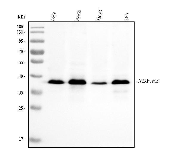
Figure 1. Western blot analysis of NDFIP2 using anti-NDFIP2 antibody (M08384).
Electrophoresis was performed on a 5-20% SDS-PAGE gel at 70V (Stacking gel) / 90V (Resolving gel) for 2-3 hours. The sample well of each lane was loaded with 30 ug of sample under reducing conditions.
Lane 1: human A549 whole cell lysates,
Lane 2: human HepG2 whole cell lysates,
Lane 3: human MCF-7 whole cell lysates,
Lane 4: human Hela whole cell lysates.
After electrophoresis, proteins were transferred to a nitrocellulose membrane at 150 mA for 50-90 minutes. Blocked the membrane with 5% non-fat milk/TBS for 1.5 hour at RT. The membrane was incubated with mouse anti-NDFIP2 antigen affinity purified monoclonal antibody (Catalog # M08384) at 0.5 μg/mL overnight at 4°C, then washed with TBS-0.1%Tween 3 times with 5 minutes each and probed with a goat anti-mouse IgG-HRP secondary antibody at a dilution of 1:10000 for 1.5 hour at RT. The signal is developed using an Enhanced Chemiluminescent detection (ECL) kit (Catalog # EK1001) with Tanon 5200 system. A specific band was detected for NDFIP2 at approximately 39 kDa. The expected band size for NDFIP2 is at 36 kDa.

Click image to see more details
Figure 2. IHC analysis of NDFIP2 using anti-NDFIP2 antibody (M08384).
NDFIP2 was detected in a paraffin-embedded section of human placenta tissue. Heat mediated antigen retrieval was performed in EDTA buffer (pH 8.0, epitope retrieval solution). The tissue section was blocked with 10% goat serum. The tissue section was then incubated with 2 μg/ml mouse anti-NDFIP2 Antibody (M08384) overnight at 4°C. Peroxidase Conjugated Goat Anti-mouse IgG was used as secondary antibody and incubated for 30 minutes at 37°C. The tissue section was developed using HRP Conjugated Mouse IgG Super Vision Assay Kit (Catalog # SV0001) with DAB as the chromogen.

Click image to see more details
Figure 3. IHC analysis of NDFIP2 using anti-NDFIP2 antibody (M08384).
NDFIP2 was detected in a paraffin-embedded section of human squamous cell lung carcinoma tissue. Heat mediated antigen retrieval was performed in EDTA buffer (pH 8.0, epitope retrieval solution). The tissue section was blocked with 10% goat serum. The tissue section was then incubated with 2 μg/ml mouse anti-NDFIP2 Antibody (M08384) overnight at 4°C. Peroxidase Conjugated Goat Anti-mouse IgG was used as secondary antibody and incubated for 30 minutes at 37°C. The tissue section was developed using HRP Conjugated Mouse IgG Super Vision Assay Kit (Catalog # SV0001) with DAB as the chromogen.

Click image to see more details
Figure 4. IHC analysis of NDFIP2 using anti-NDFIP2 antibody (M08384).
NDFIP2 was detected in a paraffin-embedded section of human breast cancer tissue. Heat mediated antigen retrieval was performed in EDTA buffer (pH 8.0, epitope retrieval solution). The tissue section was blocked with 10% goat serum. The tissue section was then incubated with 2 μg/ml mouse anti-NDFIP2 Antibody (M08384) overnight at 4°C. Peroxidase Conjugated Goat Anti-mouse IgG was used as secondary antibody and incubated for 30 minutes at 37°C. The tissue section was developed using HRP Conjugated Mouse IgG Super Vision Assay Kit (Catalog # SV0001) with DAB as the chromogen.

Click image to see more details
Figure 5. IHC analysis of NDFIP2 using anti-NDFIP2 antibody (M08384).
NDFIP2 was detected in a paraffin-embedded section of human spleen tissue. Heat mediated antigen retrieval was performed in EDTA buffer (pH 8.0, epitope retrieval solution). The tissue section was blocked with 10% goat serum. The tissue section was then incubated with 2 μg/ml mouse anti-NDFIP2 Antibody (M08384) overnight at 4°C. Peroxidase Conjugated Goat Anti-mouse IgG was used as secondary antibody and incubated for 30 minutes at 37°C. The tissue section was developed using HRP Conjugated Mouse IgG Super Vision Assay Kit (Catalog # SV0001) with DAB as the chromogen.

Click image to see more details
Figure 6. IF analysis of NDFIP2 using anti-NDFIP2 antibody (M08384).
NDFIP2 was detected in an immunocytochemical section of A431 cells. Enzyme antigen retrieval was performed using IHC enzyme antigen retrieval reagent (AR0022) for 15 mins. The cells were blocked with 10% goat serum. And then incubated with 5 μg/mL mouse anti-NDFIP2 Antibody (M08384) overnight at 4°C. DyLight®594 Conjugated Goat Anti-Mouse IgG (BA1141) was used as secondary antibody at 1:100 dilution and incubated for 30 minutes at 37°C. The section was counterstained with DAPI. Visualize using a fluorescence microscope and filter sets appropriate for the label used.

Click image to see more details
Figure 7. Flow Cytometry analysis of JK cells using anti-NDFIP2 antibody (M08384).
Overlay histogram showing JK cells stained with M08384 (Blue line). To facilitate intracellular staining, cells were fixed with 4% paraformaldehyde and permeabilized with permeabilization buffer. The cells were blocked with 10% normal goat serum. And then incubated with mouse anti-NDFIP2 Antibody (M08384, 1 μg/1x106 cells) for 30 min at 20°C. DyLight®488 conjugated goat anti-mouse IgG (BA1126, 5-10 μg/1x106 cells) was used as secondary antibody for 30 minutes at 20°C. Isotype control antibody (Green line) was mouse IgG (1 μg/1x106) used under the same conditions. Unlabelled sample without incubation with primary antibody and secondary antibody (Red line) was used as a blank control.
Protein Target Info & Infographic
Gene/Protein Information For NDFIP2 (Source: Uniprot.org, NCBI)
Gene Name
NDFIP2
Full Name
NEDD4 family-interacting protein 2
Weight
Alternative Names
FLJ25842; KIAA1165N4WBP5APutative MAPK-activating protein PM04/PM05/PM06/PM07; MAPK-activating protein PM04 PM05 PM06 PM07; N4wbp5a; Nedd4 family interacting protein 2; NEDD4 family-interacting protein 2; NEDD4 WW domain-binding protein 5A; NF-kappa-B-activating protein 413; Putative NF-kappa-B-activating protein 413 NDFIP2 N4WBP5A Nedd4 family interacting protein 2 NEDD4 family-interacting protein 2|MAPK-activating protein PM04 PM05 PM06 PM07|NEDD4 WW domain-binding protein 5A|NF-kappa-B-activating protein 413|putative MAPK-activating protein PM04/PM05/PM06/PM07|putative NF-kappa-B-activating protein 413
*If product is indicated to react with multiple species, protein info is based on the gene entry specified above in "Species".For more info on NDFIP2, check out the NDFIP2 Infographic

We have 30,000+ of these available, one for each gene! Check them out.
In this infographic, you will see the following information for NDFIP2: database IDs, superfamily, protein function, synonyms, molecular weight, chromosomal locations, tissues of expression, subcellular locations, post-translational modifications, and related diseases, research areas & pathways. If you want to see more information included, or would like to contribute to it and be acknowledged, please contact [email protected].
Specific Publications For Anti-NDFIP2 Antibody Picoband™ (monoclonal, 10D6D7) (M08384)
Hello CJ!
No publications found for M08384
*Do you have publications using this product? Share with us and receive a reward. Ask us for more details.
Recommended Resources
Here are featured tools and databases that you might find useful.
- Boster's Pathways Library
- Protein Databases
- Bioscience Research Protocol Resources
- Data Processing & Analysis Software
- Photo Editing Software
- Scientific Literature Resources
- Research Paper Management Tools
- Molecular Biology Software
- Primer Design Tools
- Bioinformatics Tools
- Phylogenetic Tree Analysis
Customer Reviews
Have you used Anti-NDFIP2 Antibody Picoband™ (monoclonal, 10D6D7)?
Submit a review and receive an Amazon gift card.
- $30 for a review with an image
0 Reviews For Anti-NDFIP2 Antibody Picoband™ (monoclonal, 10D6D7)
Customer Q&As
Have a question?
Find answers in Q&As, reviews.
Can't find your answer?
Submit your question
