Click image to see more details
Product Info Summary
| SKU: | A01106 |
|---|---|
| Size: | 100 μg/vial |
| Reactive Species: | Human, Monkey, Mouse, Rat |
| Host: | Rabbit |
| Application: | ELISA, IHC, WB |
Customers Who Bought This Also Bought

Product info
Product Name
Anti-liver Arginase/ARG1 Antibody Picoband®
View all Arginase 1/ARG1/liver Arginase Antibodies
SKU/Catalog Number
A01106
Size
100 μg/vial
Form
Lyophilized
Description
Boster Bio Anti-liver Arginase/ARG1 Antibody Picoband® catalog # A01106. Tested in ELISA, IHC, WB applications. This antibody reacts with Human, Monkey, Mouse, Rat. The brand Picoband indicates this is a premium antibody that guarantees superior quality, high affinity, and strong signals with minimal background in Western blot applications. Only our best-performing antibodies are designated as Picoband, ensuring unmatched performance.
Storage & Handling
Store at -20˚C for one year from date of receipt. After reconstitution, at 4˚C for one month. It can also be aliquotted and stored frozen at -20˚C for six months. Avoid repeated freeze-thaw cycles.
Cite This Product
Anti-liver Arginase/ARG1 Antibody Picoband® (Boster Biological Technology, Pleasanton CA, USA, Catalog # A01106)
Host
Rabbit
Contents
Each vial contains 4mg Trehalose, 0.9mg NaCl and 0.2mg Na2HPO4.
Clonality
Polyclonal
Isotype
Rabbit IgG
Immunogen
E.coli-derived human liver Arginase/ARG1 recombinant protein (Position: E25-D183).
*Blocking peptide can be purchased. Costs vary based on immunogen length. Contact us for pricing.
Cross-reactivity
No cross-reactivity with other proteins.
Reactive Species
A01106 is reactive to ARG1 in Human, Monkey, Mouse, Rat
Reconstitution
Add 0.2ml of distilled water will yield a concentration of 500ug/ml.
Observed Molecular Weight
37 kDa
Calculated molecular weight
51735 MW
Background of Arginase 1/ARG1/liver Arginase
ARG1 (arginase, live) is a cytosolic enzyme and expressed predominantly in the liver as a component of the urea cycle. The isoform encoded by ARG1, referred to as the liver, or A-I, isoform, contributes 98% of the arginase activity in liver but is also present in red cells. Using a rat liver ARG1 cDNA clone to probe a human liver cDNA library, Haraguchi et al. (1987) isolated and characterized a cDNA corresponding to the ARG1 gene. The ARG1 gene is mapped on 6q23.2 and the arginase gene contains 8 exons. By immunologic studies, 90% of the arginase in red blood cell and liver was precipitated by the antibody, whereas only 50% of the arginase in kidney, brain, and the gastrointestinal tract reacted with it. Inherited deficiency of this enzyme results in argininemia, an autosomal recessive disorder characterized by hyperammonemia. Two transcript variants encoding different isoforms have been found for this gene.
Antibody Validation
Boster validates all antibodies on WB, IHC, ICC, Immunofluorescence, and ELISA with known positive control and negative samples to ensure specificity and high affinity, including thorough antibody incubations.
Application & Images
Applications
A01106 is guaranteed for ELISA, IHC, WB Boster Guarantee
Assay Dilutions Recommendation
The recommendations below provide a starting point for assay optimization. The actual working concentration varies and should be decided by the user.
Western blot, 0.25-0.5 μg/ml
Immunohistochemistry(Paraffin-embedded Section), 2-5 μg/ml
ELISA, 0.1-0.5 μg/ml
Positive Control
WB: human HCCP tissue, monkey liver tissue, rat liver tissue, mouse liver tissue, rat testis tissue
IHC: human liver cancer tissue, mouse liver tissue, rat liver tissue
Validation Images & Assay Conditions
Click image to see more details
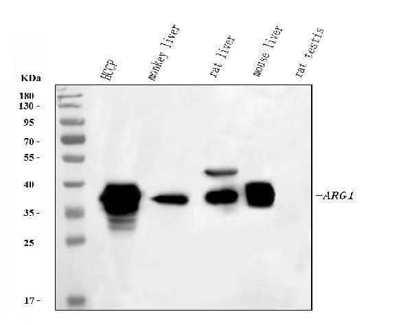
Figure 1. Western blot analysis of ARG1 using anti-ARG1 antibody (A01106).
Electrophoresis was performed on a 5-20% SDS-PAGE gel at 70V (Stacking gel) / 90V (Resolving gel) for 2-3 hours. The sample well of each lane was loaded with 30 ug of sample under reducing conditions.
Lane 1: human HCCP tissue lysates,
Lane 2: monkey liver tissue lysates,
Lane 3: rat liver tissue lysates,
Lane 4: mouse liver tissue lysates,
Lane 5: rat testis tissue lysates.
After electrophoresis, proteins were transferred to a nitrocellulose membrane at 150 mA for 50-90 minutes. Blocked the membrane with 5% non-fat milk/TBS for 1.5 hour at RT. The membrane was incubated with rabbit anti-ARG1 antigen affinity purified polyclonal antibody (Catalog # A01106) at 0.5 μg/mL overnight at 4°C, then washed with TBS-0.1%Tween 3 times with 5 minutes each and probed with a goat anti-rabbit IgG-HRP secondary antibody at a dilution of 1:5000 for 1.5 hour at RT. The signal is developed using an Enhanced Chemiluminescent detection (ECL) kit (Catalog # EK1002) with Tanon 5200 system. A specific band was detected for ARG1 at approximately 37 kDa. The expected band size for ARG1 is at 35 kDa.

Click image to see more details
Figure 2. IHC analysis of ARG1 using anti-ARG1 antibody (A01106).
ARG1 was detected in a paraffin-embedded section of human liver cancer tissue. Heat mediated antigen retrieval was performed in EDTA buffer (pH 8.0, epitope retrieval solution). The tissue section was blocked with 10% goat serum. The tissue section was then incubated with 2 μg/ml rabbit anti-ARG1 Antibody (A01106) overnight at 4°C. Biotinylated goat anti-rabbit IgG was used as secondary antibody and incubated for 30 minutes at 37°C. The tissue section was developed using Strepavidin-Biotin-Complex (SABC) (Catalog # SA1022) with DAB as the chromogen.

Click image to see more details
Figure 3. IHC analysis of ARG1 using anti-ARG1 antibody (A01106).
ARG1 was detected in a paraffin-embedded section of mouse liver tissue. Heat mediated antigen retrieval was performed in EDTA buffer (pH 8.0, epitope retrieval solution). The tissue section was blocked with 10% goat serum. The tissue section was then incubated with 2 μg/ml rabbit anti-ARG1 Antibody (A01106) overnight at 4°C. Biotinylated goat anti-rabbit IgG was used as secondary antibody and incubated for 30 minutes at 37°C. The tissue section was developed using Strepavidin-Biotin-Complex (SABC) (Catalog # SA1022) with DAB as the chromogen.

Click image to see more details
Figure 4. IHC analysis of ARG1 using anti-ARG1 antibody (A01106).
ARG1 was detected in a paraffin-embedded section of rat liver tissue. Heat mediated antigen retrieval was performed in EDTA buffer (pH 8.0, epitope retrieval solution). The tissue section was blocked with 10% goat serum. The tissue section was then incubated with 2 μg/ml rabbit anti-ARG1 Antibody (A01106) overnight at 4°C. Biotinylated goat anti-rabbit IgG was used as secondary antibody and incubated for 30 minutes at 37°C. The tissue section was developed using Strepavidin-Biotin-Complex (SABC) (Catalog # SA1022) with DAB as the chromogen.
Protein Target Info & Infographic
Gene/Protein Information For ARG1 (Source: Uniprot.org, NCBI)
Gene Name
ARG1
Full Name
Arginase-1
Weight
51735 MW
Superfamily
arginase family
Alternative Names
Arginase-1; Liver-type arginase; Type I arginase; ARG1 ARG1 arginase 1 arginase-1|arginase, liver|liver-type arginase|type I arginase
*If product is indicated to react with multiple species, protein info is based on the gene entry specified above in "Species".For more info on ARG1, check out the ARG1 Infographic

We have 30,000+ of these available, one for each gene! Check them out.
In this infographic, you will see the following information for ARG1: database IDs, superfamily, protein function, synonyms, molecular weight, chromosomal locations, tissues of expression, subcellular locations, post-translational modifications, and related diseases, research areas & pathways. If you want to see more information included, or would like to contribute to it and be acknowledged, please contact [email protected].
Specific Publications For Anti-liver Arginase/ARG1 Antibody Picoband® (A01106)
Hello CJ!
A01106 has been cited in 3 publications:
*The publications in this section are manually curated by our staff scientists. They may differ from Bioz's machine gathered results. Both are accurate. If you find a publication citing this product but is missing from this list, please let us know we will issue you a thank-you coupon.
Liao H,Li Y,Zhang X,Zhao X,Zheng D,Shen D,Li R. Protective Effects of Thalidomide on High-Glucose-Induced Podocyte Injury through In Vitro Modulation of Macrophage M1/M2 Differentiation.J Immunol Res.2020 Aug 27;2020:8263598.doi:10.1155/2020/ 8263598.PMID
Species: Mouse
A01106 usage in article: APP:WB, SAMPLE:MACROPHAGES, DILUTION:1:500
Panax notoginseng Saponins Regulate Macrophage Polarization under Hyperglycemic Condition via NF-%u03BAB Signaling Pathway
CFTR protects against vascular inflammation and atherogenesis in apolipoprotein E-deficient mice
Recommended Resources
Here are featured tools and databases that you might find useful.
- Boster's Pathways Library
- Protein Databases
- Bioscience Research Protocol Resources
- Data Processing & Analysis Software
- Photo Editing Software
- Scientific Literature Resources
- Research Paper Management Tools
- Molecular Biology Software
- Primer Design Tools
- Bioinformatics Tools
- Phylogenetic Tree Analysis
Customer Reviews
Have you used Anti-liver Arginase/ARG1 Antibody Picoband®?
Submit a review and receive an Amazon gift card.
- $30 for a review with an image
0 Reviews For Anti-liver Arginase/ARG1 Antibody Picoband®
Customer Q&As
Have a question?
Find answers in Q&As, reviews.
Can't find your answer?
Submit your question
5 Customer Q&As for Anti-liver Arginase/ARG1 Antibody Picoband®
Question
Our lab were satisfied with the WB result of your anti-liver Arginase/ARG1 antibody. However we have seen positive staining in liver cytoplasm. using this antibody. Is that expected? Could you tell me where is ARG1 supposed to be expressed?
Verified Customer
Verified customer
Asked: 2020-02-27
Answer
From what I have seen in literature, liver does express ARG1. Generally ARG1 expresses in cytoplasm. Regarding which tissues have ARG1 expression, here are a few articles citing expression in various tissues:
Blood, Pubmed ID: 3174433
Erythroblast, Pubmed ID: 14574404
Liver, Pubmed ID: 2241902, 3540966, 24275569
Liver, and Skeletal muscle, Pubmed ID: 15489334
Boster Scientific Support
Answered: 2020-02-27
Question
We have been able to see staining in human blood. Any tips? Is anti-liver Arginase/ARG1 antibody supposed to stain blood positively?
Verified Customer
Verified customer
Asked: 2020-01-20
Answer
Based on literature blood does express ARG1. Based on Uniprot.org, ARG1 is expressed in liver, blood, erythroblast, liver skeletal muscle, among other tissues. Regarding which tissues have ARG1 expression, here are a few articles citing expression in various tissues:
Blood, Pubmed ID: 3174433
Erythroblast, Pubmed ID: 14574404
Liver, Pubmed ID: 2241902, 3540966, 24275569
Liver, and Skeletal muscle, Pubmed ID: 15489334
Boster Scientific Support
Answered: 2020-01-20
Question
We need using your anti-liver Arginase/ARG1 antibody for response to selenium ion studies. Has this antibody been tested with western blotting on rat liver tissue? We would like to see some validation images before ordering.
Verified Customer
Verified customer
Asked: 2019-11-27
Answer
I appreciate your inquiry. This A01106 anti-liver Arginase/ARG1 antibody is tested on rat liver tissue. It is guaranteed to work for ELISA, WB in human, mouse, rat. Our Boster guarantee will cover your intended experiment even if the sample type has not been be directly tested.
Boster Scientific Support
Answered: 2019-11-27
Question
We are currently using anti-liver Arginase/ARG1 antibody A01106 for rat tissue, and we are happy with the WB results. The species of reactivity given in the datasheet says human, mouse, rat. Is it true that the antibody can work on zebrafish tissues as well?
Verified Customer
Verified customer
Asked: 2019-10-29
Answer
The anti-liver Arginase/ARG1 antibody (A01106) has not been validated for cross reactivity specifically with zebrafish tissues, though there is a good chance of cross reactivity. We have an innovator award program that if you test this antibody and show it works in zebrafish you can get your next antibody for free. Please contact me if I can help you with anything.
Boster Scientific Support
Answered: 2019-10-29
Question
Our lab used your anti-liver Arginase/ARG1 antibody for ELISA on liver skeletal muscle in a previous experiment. I am using rat, and I plan to use the antibody for WB next. We are interested in examining liver skeletal muscle as well as erythroblast in our next experiment. Could you please give me some suggestion on which antibody would work the best for WB?
Verified Customer
Verified customer
Asked: 2019-06-06
Answer
I looked at the website and datasheets of our anti-liver Arginase/ARG1 antibody and it seems that A01106 has been validated on rat in both ELISA and WB. Thus A01106 should work for your application. Our Boster satisfaction guarantee will cover this product for WB in rat even if the specific tissue type has not been validated. We do have a comprehensive range of products for WB detection and you can check out our website bosterbio.com to find out more information about them.
Boster Scientific Support
Answered: 2019-06-06
