Click image to see more details
Product Info Summary
| SKU: | A01373-1 |
|---|---|
| Size: | 100 μg/vial |
| Reactive Species: | Human |
| Host: | Rabbit |
| Application: | ELISA, IHC, WB |
Customers Who Bought This Also Bought

Product info
Product Name
Anti-IGFBP2 Antibody Picoband™
SKU/Catalog Number
A01373-1
Size
100 μg/vial
Form
Lyophilized
Description
Boster Bio Anti-IGFBP2 Antibody Picoband™ catalog # A01373-1. Tested in ELISA, IHC, WB applications. This antibody reacts with Human.
Storage & Handling
Store at -20˚C for one year from date of receipt. After reconstitution, at 4˚C for one month. It can also be aliquotted and stored frozen at -20˚C for six months. Avoid repeated freeze-thaw cycles.
Cite This Product
Anti-IGFBP2 Antibody Picoband™ (Boster Biological Technology, Pleasanton CA, USA, Catalog # A01373-1)
Host
Rabbit
Contents
Each vial contains 4mg Trehalose, 0.9mg NaCl, 0.2mg Na2HPO4, 0.05mg NaN3.
Clonality
Polyclonal
Isotype
Rabbit IgG
Immunogen
E. coli-derived human IGFBP2 recombinant protein (Position: A36-Q325).
*Blocking peptide can be purchased. Costs vary based on immunogen length. Contact us for pricing.
Cross-reactivity
No cross-reactivity with other proteins.
Reactive Species
A01373-1 is reactive to IGFBP2 in Human
Applications
A01373-1 is guaranteed for ELISA, IHC, WB Boster Guarantee
Observed Molecular Weight
35 kDa
Calculated molecular weight
Background of IGFBP-2
The superfamily of insulin-like growth factor (IGF) binding proteins include the six high-affinity IGF binding proteins (IGFBP) and at least four additional low-affinity binding proteins referred to as IGFBP related proteins (IGFBP-rP). All IGFBP superfamily members are cysteine-rich proteins with conserved cysteine residues, which are clustered in the amino- and carboxy-terminal thirds of the molecule. IGFBPs modulate the biological activities of IGF proteins. Some IGFBPs may also have intrinsic bioactivity that is independent of their ability to bind IGF proteins. Post-translational modifications of IGFBPs, including glycosylation, phosphorylation and proteolysis, have been shown to modify the affinities of the binding proteins to IGF. Human IGFBP-2 cDNA encodes a 328 amino acid (aa) residue precursor protein with a putative 39 aa residue signal peptide that is processed to generate the 289 aa residue mature protein. IGFBP-2 contains an integrin receptor recognition sequence (RGD sequence) but lacks potential N-linked glycosylation sites. During development, IGFBP-2 is expressed in a number of tissues. The highest expression level is found in the central nervous system. In adults, high expression levels are also detected in the central nervous system and in a number of reproductive tissues. IGFBP-2 binds preferentially to IGF II, exhibiting a 2-10 fold higher affinity for IGF II than for IGF I.
Antibody Validation
Boster validates all antibodies on WB, IHC, ICC, Immunofluorescence, and ELISA with known positive control and negative samples to ensure specificity and high affinity, including thorough antibody incubations.
Assay dilution & Images
Reconstitution
Add 0.2ml of distilled water will yield a concentration of 500ug/ml.
Assay Dilutions Recommendation
The recommendations below provide a starting point for assay optimization. The actual working concentration varies and should be decided by the user.
Western blot, 0.1-0.5μg/ml, Human
Immunohistochemistry (Paraffin-embedded Section), 0.5-1μg/ml, Human
ELISA (Cap), 1-5μg/ml, Human
Validation Images & Assay Conditions
Click image to see more details
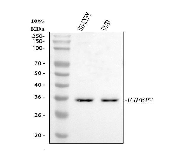
Figure 1. Western blot analysis of IGFBP2 using anti-IGFBP2 antibody (A01373-1).
Electrophoresis was performed on a 5-20% SDS-PAGE gel at 70V (Stacking gel) / 90V (Resolving gel) for 2-3 hours. The sample well of each lane was loaded with 30 ug of sample under reducing conditions.
Lane 1: human SH-SY5Y whole cell lysates,
Lane 2: human T-47D whole cell lysates.
After electrophoresis, proteins were transferred to a nitrocellulose membrane at 150 mA for 50-90 minutes. Blocked the membrane with 5% non-fat milk/TBS for 1.5 hour at RT. The membrane was incubated with rabbit anti-IGFBP2 antigen affinity purified polyclonal antibody (Catalog # A01373-1) at 0.5 μg/mL overnight at 4°C, then washed with TBS-0.1%Tween 3 times with 5 minutes each and probed with a goat anti-rabbit IgG-HRP secondary antibody at a dilution of 1:5000 for 1.5 hour at RT. The signal is developed using an Enhanced Chemiluminescent detection (ECL) kit (Catalog # EK1002) with Tanon 5200 system. A specific band was detected for IGFBP2 at approximately 35 kDa. The expected band size for IGFBP2 is at 35 kDa.

Click image to see more details
Figure 2. IHC analysis of IGFBP2 using anti-IGFBP2 antibody (A01373-1).
IGFBP2 was detected in paraffin-embedded section of human lung cancer tissue. Heat mediated antigen retrieval was performed in citrate buffer (pH6, epitope retrieval solution) for 20 mins. The tissue section was blocked with 10% goat serum. The tissue section was then incubated with 1ug/ml rabbit anti-IGFBP2 Antibody (A01373-1) overnight at 4 Biotinylated goat anti-rabbit IgG was used as secondary antibody and incubated for 30 minutes at 37 The tissue section was developed using Strepavidin-Biotin-Complex (SABC)(Catalog # SA1022) with DAB as the chromogen.

Click image to see more details
Figure 3. IHC analysis of IGFBP2 using anti-IGFBP2 antibody (A01373-1).
IGFBP2 was detected in paraffin-embedded section of human mammary cancer tissue. Heat mediated antigen retrieval was performed in citrate buffer (pH6, epitope retrieval solution) for 20 mins. The tissue section was blocked with 10% goat serum. The tissue section was then incubated with 1ug/ml rabbit anti-IGFBP2 Antibody (A01373-1) overnight at 4 Biotinylated goat anti-rabbit IgG was used as secondary antibody and incubated for 30 minutes at 37 The tissue section was developed using Strepavidin-Biotin-Complex (SABC)(Catalog # SA1022) with DAB as the chromogen.

Click image to see more details
Figure 4. Sandwich ELISA - Recombinant human IGFBP2 protein standard curve.
Use in combination with reagents from Human IGFBP2 ELISA Kit EZ-Set (DIY Antibody Pairs) (EZ0384).
Protein Target Info & Infographic
Gene/Protein Information For IGFBP2 (Source: Uniprot.org, NCBI)
Gene Name
IGFBP2
Full Name
Insulin-like growth factor-binding protein 2
Weight
Alternative Names
BP2; IBP2; IBP-2; IGF-binding protein 2; IGFBP2; IGFBP-2; IGF-BP53; insulin-like growth factor binding protein 2 (36kD); insulin-like growth factor binding protein 2, 36kDa; insulin-like growth factor-binding protein 2 IGFBP2 IBP2, IGF-BP53 insulin like growth factor binding protein 2 insulin-like growth factor-binding protein 2|IGF-binding protein 2|insulin-like growth factor binding protein 2, 36kDa
*If product is indicated to react with multiple species, protein info is based on the gene entry specified above in "Species".For more info on IGFBP2, check out the IGFBP2 Infographic

We have 30,000+ of these available, one for each gene! Check them out.
In this infographic, you will see the following information for IGFBP2: database IDs, superfamily, protein function, synonyms, molecular weight, chromosomal locations, tissues of expression, subcellular locations, post-translational modifications, and related diseases, research areas & pathways. If you want to see more information included, or would like to contribute to it and be acknowledged, please contact [email protected].
Specific Publications For Anti-IGFBP2 Antibody Picoband™ (A01373-1)
Hello CJ!
No publications found for A01373-1
*Do you have publications using this product? Share with us and receive a reward. Ask us for more details.
Recommended Resources
Here are featured tools and databases that you might find useful.
- Boster's Pathways Library
- Protein Databases
- Bioscience Research Protocol Resources
- Data Processing & Analysis Software
- Photo Editing Software
- Scientific Literature Resources
- Research Paper Management Tools
- Molecular Biology Software
- Primer Design Tools
- Bioinformatics Tools
- Phylogenetic Tree Analysis
Customer Reviews
Have you used Anti-IGFBP2 Antibody Picoband™?
Submit a review and receive an Amazon gift card.
- $30 for a review with an image
0 Reviews For Anti-IGFBP2 Antibody Picoband™
Customer Q&As
Have a question?
Find answers in Q&As, reviews.
Can't find your answer?
Submit your question
