Click image to see more details
-

-
-
-
-
+3
Product Info Summary
| SKU: | A05257-2 |
|---|---|
| Size: | 100 µg/vial |
| Reactive Species: | Human, Mouse |
| Host: | Rabbit |
| Application: | ELISA, IHC, WB |
Customers Who Bought This Also Bought


Product info
Product Name
Anti-HOXD10 Antibody Picoband™
SKU/Catalog Number
A05257-2
Size
100 µg/vial
Form
Lyophilized
Description
Boster Bio Anti-HOXD10 Antibody Picoband™ catalog # A05257-2. Tested in ELISA, IHC, WB applications. This antibody reacts with Human, Mouse.
Storage & Handling
At -20°C for one year from date of receipt. After reconstitution, at 4°C for one month. It can also be aliquotted and stored frozen at -20°C for six months. Avoid repeated freezing and thawing.
Cite This Product
Anti-HOXD10 Antibody Picoband™ (Boster Biological Technology, Pleasanton CA, USA, Catalog # A05257-2)
Host
Rabbit
Contents
Each vial contains 4 mg Trehalose, 0.9 mg NaCl, 0.2 mg Na2HPO4.
Clonality
Polyclonal
Isotype
Rabbit IgG
Immunogen
E.coli-derived human HOXD10 recombinant protein (Position: A9-K252).
*Blocking peptide can be purchased. Costs vary based on immunogen length. Contact us for pricing.
Cross-reactivity
No cross-reactivity with other proteins.
Reactive Species
A05257-2 is reactive to HOXD10 in Human, Mouse
Applications
A05257-2 is guaranteed for ELISA, IHC, WB Boster Guarantee
Observed Molecular Weight
40 kDa
Calculated molecular weight
39411 MW
Background of HOXD10
Homeobox D10, also known as HOXD10, is a protein which in humans is encoded by the HOXD10 gene. This gene is a member of the Abd-B homeobox family and encodes a protein with a homeobox DNA-binding domain. It is included in a cluster of homeobox D genes located on chromosome 2. The encoded nuclear protein functions as a sequence-specific transcription factor that is expressed in the developing limb buds and is involved in differentiation and limb development. Mutations in this gene have been associated with Wilm's tumor and congenital vertical talus (also known as "rocker-bottom foot" deformity or congenital convex pes valgus) and/or a foot deformity resembling that seen in Charcot-Marie-Tooth disease.
Antibody Validation
Boster validates all antibodies on WB, IHC, ICC, Immunofluorescence, and ELISA with known positive control and negative samples to ensure specificity and high affinity, including thorough antibody incubations.
Assay dilution & Images
Reconstitution
Adding 0.2 ml of distilled water will yield a concentration of 500 µg/ml.
Assay Dilutions Recommendation
The recommendations below provide a starting point for assay optimization. The actual working concentration varies and should be decided by the user.
Western blot, 0.25-0.5 µg/ml, Mouse
Immunohistochemistry(Paraffin-embedded Section), 2-5 µg/ml, Human
Direct ELISA, 0.1-0.5 µg/ml, Human
Validation Images & Assay Conditions
Click image to see more details
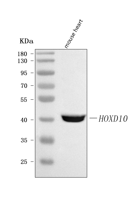
Figure 1. Western blot analysis of HOXD10 using anti-HOXD10 antibody (A05257-2).
Electrophoresis was performed on a 5-20% SDS-PAGE gel at 70V (Stacking gel) / 90V (Resolving gel) for 2-3 hours. The sample well of each lane was loaded with 30 ug of sample under reducing conditions.
Lane 1: mouse heart tissue lysates.
After electrophoresis, proteins were transferred to a nitrocellulose membrane at 150 mA for 50-90 minutes. Blocked the membrane with 5% non-fat milk/TBS for 1.5 hour at RT. The membrane was incubated with rabbit anti-HOXD10 antigen affinity purified polyclonal antibody (Catalog # A05257-2) at 0.5 μg/mL overnight at 4°C, then washed with TBS-0.1%Tween 3 times with 5 minutes each and probed with a goat anti-rabbit IgG-HRP secondary antibody at a dilution of 1:5000 for 1.5 hour at RT. The signal is developed using an Enhanced Chemiluminescent detection (ECL) kit (Catalog # EK1002) with Tanon 5200 system. A specific band was detected for HOXD10 at approximately 40 kDa. The expected band size for HOXD10 is at 38 kDa.

Click image to see more details
Figure 2. IHC analysis of HOXD10 using anti-HOXD10 antibody (A05257-2).
HOXD10 was detected in a paraffin-embedded section of human lung adenocarcinoma tissue. Heat mediated antigen retrieval was performed in EDTA buffer (pH 8.0, epitope retrieval solution). The tissue section was blocked with 10% goat serum. The tissue section was then incubated with 2 μg/ml rabbit anti-HOXD10 Antibody (A05257-2) overnight at 4°C. Peroxidase Conjugated Goat Anti-rabbit IgG was used as secondary antibody and incubated for 30 minutes at 37°C. The tissue section was developed using HRP Conjugated Rabbit IgG Super Vision Assay Kit (Catalog # SV0002) with DAB as the chromogen.

Click image to see more details
Figure 3. IHC analysis of HOXD10 using anti-HOXD10 antibody (A05257-2).
HOXD10 was detected in a paraffin-embedded section of human mucinous adenoma of ovary tissue. Heat mediated antigen retrieval was performed in EDTA buffer (pH 8.0, epitope retrieval solution). The tissue section was blocked with 10% goat serum. The tissue section was then incubated with 2 μg/ml rabbit anti-HOXD10 Antibody (A05257-2) overnight at 4°C. Peroxidase Conjugated Goat Anti-rabbit IgG was used as secondary antibody and incubated for 30 minutes at 37°C. The tissue section was developed using HRP Conjugated Rabbit IgG Super Vision Assay Kit (Catalog # SV0002) with DAB as the chromogen.

Click image to see more details
Figure 4. IHC analysis of HOXD10 using anti-HOXD10 antibody (A05257-2).
HOXD10 was detected in a paraffin-embedded section of human rectum adenocarcinoma tissue. Heat mediated antigen retrieval was performed in EDTA buffer (pH 8.0, epitope retrieval solution). The tissue section was blocked with 10% goat serum. The tissue section was then incubated with 2 μg/ml rabbit anti-HOXD10 Antibody (A05257-2) overnight at 4°C. Peroxidase Conjugated Goat Anti-rabbit IgG was used as secondary antibody and incubated for 30 minutes at 37°C. The tissue section was developed using HRP Conjugated Rabbit IgG Super Vision Assay Kit (Catalog # SV0002) with DAB as the chromogen.

Click image to see more details
Figure 5. IHC analysis of HOXD10 using anti-HOXD10 antibody (A05257-2).
HOXD10 was detected in a paraffin-embedded section of human thyroid papillary carcinoma tissue. Heat mediated antigen retrieval was performed in EDTA buffer (pH 8.0, epitope retrieval solution). The tissue section was blocked with 10% goat serum. The tissue section was then incubated with 2 μg/ml rabbit anti-HOXD10 Antibody (A05257-2) overnight at 4°C. Peroxidase Conjugated Goat Anti-rabbit IgG was used as secondary antibody and incubated for 30 minutes at 37°C. The tissue section was developed using HRP Conjugated Rabbit IgG Super Vision Assay Kit (Catalog # SV0002) with DAB as the chromogen.

Click image to see more details
Figure 6. IHC analysis of HOXD10 using anti-HOXD10 antibody (A05257-2).
HOXD10 was detected in a paraffin-embedded section of human tonsil tissue. Heat mediated antigen retrieval was performed in EDTA buffer (pH 8.0, epitope retrieval solution). The tissue section was blocked with 10% goat serum. The tissue section was then incubated with 2 μg/ml rabbit anti-HOXD10 Antibody (A05257-2) overnight at 4°C. Peroxidase Conjugated Goat Anti-rabbit IgG was used as secondary antibody and incubated for 30 minutes at 37°C. The tissue section was developed using HRP Conjugated Rabbit IgG Super Vision Assay Kit (Catalog # SV0002) with DAB as the chromogen.

Click image to see more details
Figure 7. IHC analysis of HOXD10 using anti-HOXD10 antibody (A05257-2).
HOXD10 was detected in a paraffin-embedded section of human urothelial carcinoma tissue. Heat mediated antigen retrieval was performed in EDTA buffer (pH 8.0, epitope retrieval solution). The tissue section was blocked with 10% goat serum. The tissue section was then incubated with 2 μg/ml rabbit anti-HOXD10 Antibody (A05257-2) overnight at 4°C. Peroxidase Conjugated Goat Anti-rabbit IgG was used as secondary antibody and incubated for 30 minutes at 37°C. The tissue section was developed using HRP Conjugated Rabbit IgG Super Vision Assay Kit (Catalog # SV0002) with DAB as the chromogen.
Protein Target Info & Infographic
Gene/Protein Information For HOXD10 (Source: Uniprot.org, NCBI)
Gene Name
HOXD10
Full Name
Homeobox protein Hox-D10
Weight
39411 MW
Superfamily
Abd-B homeobox family
Alternative Names
homeo box 4D; homeo box D10; homeobox D10; Homeobox protein Hox-4D; Homeobox protein Hox-4E; homeobox protein Hox-D10; Hox-4.4; HOX4DHOX4; HOX4E HOXD10 HOX4, HOX4D, HOX4E, Hox-4.4 homeobox D10 homeobox protein Hox-D10|homeo box 4D|homeo box D10|homeobox protein Hox-4D|homeobox protein Hox-4E
*If product is indicated to react with multiple species, protein info is based on the gene entry specified above in "Species".For more info on HOXD10, check out the HOXD10 Infographic

We have 30,000+ of these available, one for each gene! Check them out.
In this infographic, you will see the following information for HOXD10: database IDs, superfamily, protein function, synonyms, molecular weight, chromosomal locations, tissues of expression, subcellular locations, post-translational modifications, and related diseases, research areas & pathways. If you want to see more information included, or would like to contribute to it and be acknowledged, please contact [email protected].
Specific Publications For Anti-HOXD10 Antibody Picoband™ (A05257-2)
Hello CJ!
No publications found for A05257-2
*Do you have publications using this product? Share with us and receive a reward. Ask us for more details.
Recommended Resources
Here are featured tools and databases that you might find useful.
- Boster's Pathways Library
- Protein Databases
- Bioscience Research Protocol Resources
- Data Processing & Analysis Software
- Photo Editing Software
- Scientific Literature Resources
- Research Paper Management Tools
- Molecular Biology Software
- Primer Design Tools
- Bioinformatics Tools
- Phylogenetic Tree Analysis
Customer Reviews
Have you used Anti-HOXD10 Antibody Picoband™?
Submit a review and receive an Amazon gift card.
- $30 for a review with an image
0 Reviews For Anti-HOXD10 Antibody Picoband™
Customer Q&As
Have a question?
Find answers in Q&As, reviews.
Can't find your answer?
Submit your question
